Method and system for full-field interference microscopy imaging

A technology of interference microscopy, an imaging method, applied in the field of cell and intracellular imaging

Pending Publication Date: 2018-02-27
エルエルテックマネージメント
View PDF5 Cites 11 Cited by
  • Summary
  • Abstract
  • Description
  • Claims
  • Application Information

AI Technical Summary

Problems solved by technology

Finally, these techniques are not suitable for perioperative use, as several days are often required before tissue observation is possible given the different steps involved in preparing the tissue

Method used


Image

  • Method and system for full-field interference microscopy imaging
  • Method and system for full-field interference microscopy imaging
  • Method and system for full-field interference microscopy imaging

Examples


Embodiment Construction

[0060] imaging system

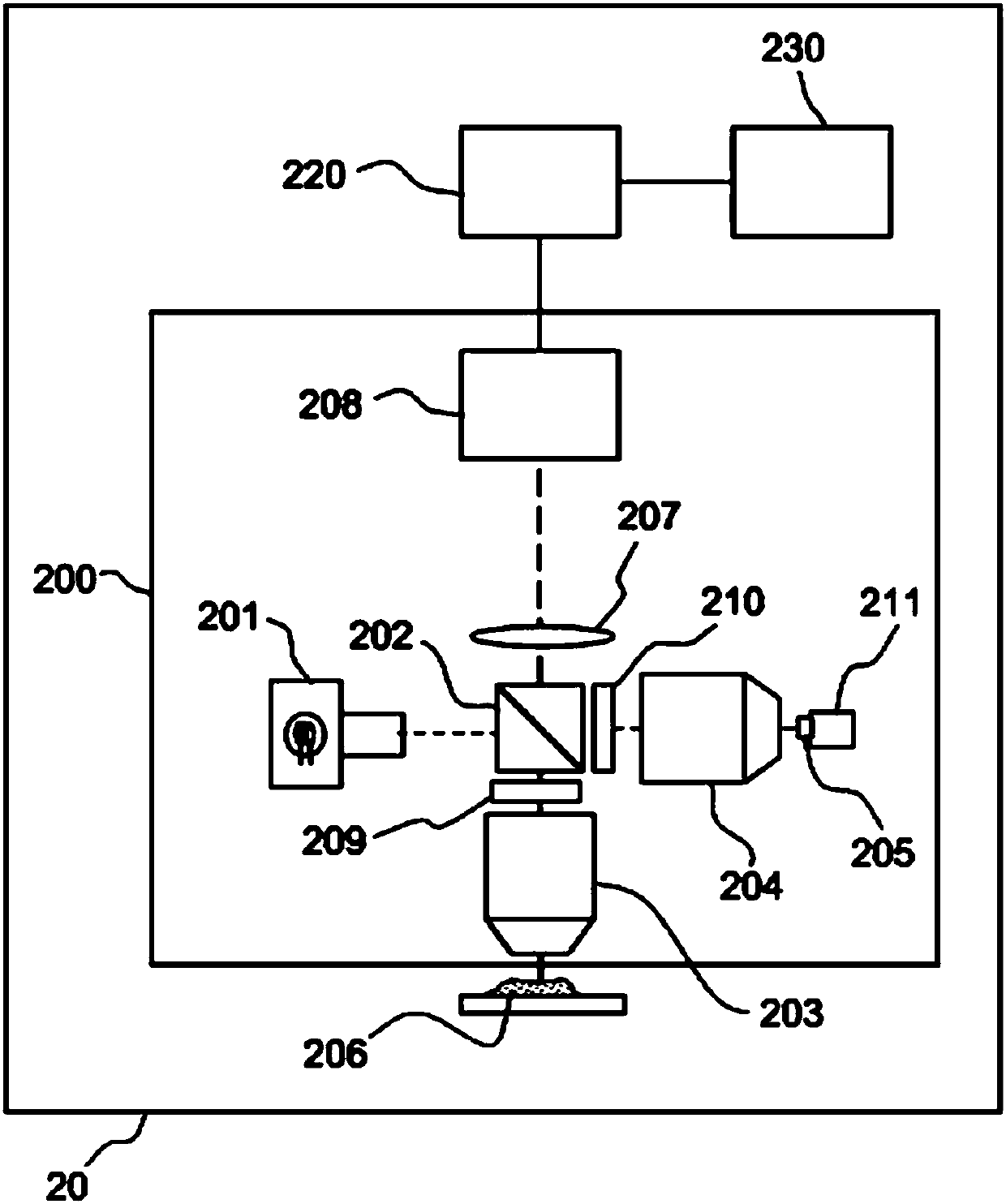
[0061] exist figure 2 An embodiment of an imaging system 20 suitable for implementing a method of imaging a three-dimensional sample according to the present description is schematically shown in FIG.

[0062] The imaging system 20 comprises an interferometric device 200 , an acquisition device 208 and at least one processing unit 220 .

[0063] The interference device 200 is adapted to generate optical interference of a reference wave and an object wave. On the one hand, the reference wave is obtained by reflecting the spatially incoherent light emitted by the light source 201 and having a low coherence length by each basic surface of the reflective surface 205 of the reference arm of the interference device; on the other hand, through the sample 206 in the depth direction Object waves are obtained by backscattering light emitted by the same source at each voxel of the slice, the sample 206 being arranged on the target arm of the interferometric d...


PUM


Abstract

The invention relates to a system (20) for full-field interference microscopy imaging of a three-dimensional diffusing sample (206). Said system includes: an interference device (200) including a reference arm on which a reflective surface (205) is arranged, the interference device being suitable for producing, at each point of an imaging field when the sample is placed on a target arm of the interference device, interference between a reference wave, obtained by reflection of incident light waves onto a basic surface of the reflective surface (205) corresponding to said point of the imaging field, and a target wave obtained by backscattering of incident light waves by means of a voxel of a slice of the sample at a given depth, said voxel corresponding to said point of the imaging field; an acquisition device (208) suitable for acquiring, at a fixed path length difference between the target arm and the reference arm, a temporal series of N two-dimensional interferometric signals resulting from the interference produced at each point of the imaging field; and a processing unit (220) configured to calculate an image (IB, IC) representing temporal variations in intensity between saidN two-dimensional interferometric signals.

Description

technical field [0001] This specification relates to a method and system for full-field interferometric microscopy imaging, which is particularly suitable for imaging cells and intracellularly. Background technique [0002] Known by the name full-field OCT (OCT is an acronym for "Optical Coherence Tomography"), the technique of acquiring images by full-field interference microscopy of incoherent light is a very Invasive, non-destructive and endogenous methods are very powerful in acquiring images of biological tissues. [0003] Full-field OCT imaging techniques are described, for example, in the article "Full-field optical coherencetomography" by A. Dubois and C. Boccara, taken from "Optical Coherence Tomography-Technology and Applications"-Wolfgang Drexler-James G.Fujimoto-Editors -Springer 2009. Full-field OCT imaging techniques are also described in French patent application FR2817030. [0004] Full-field OCT imaging techniques are based on the use of light backscatter...

Claims


Application Information

Patent Timeline
Patent Type & Authority Applications(China)
IPC IPC(8): G01N21/47G01B9/02G02B21/00G02B21/36
CPCG01B9/02091G01N21/4795G02B21/0004G02B21/365G02B21/14G02B21/18G01N21/47G01N2021/4709G02B21/082G02B21/125G06T1/0007G06T5/50G06T2207/10101
Inventor A·C·博卡拉F·哈姆斯
Owner エルエルテックマネージメント
Why Eureka
Try Eureka