Medical image processing method and medical image processing system

A medical image and processing method technology, applied in the field of image processing, can solve the problems of low accuracy and slow speed of the lesion detection method, and achieve the effect of improving the detection accuracy, speed and speed.

Active Publication Date: 2018-04-20
SHANGHAI UNITED IMAGING HEALTHCARE
View PDF7 Cites 43 Cited by
  • Summary
  • Abstract
  • Description
  • Claims
  • Application Information

AI Technical Summary

Problems solved by technology

[0004] Embodiments of the present invention provide a medical image processing method and a medical image processing system, which solve the problems of low accuracy and slow speed of existing focus detection methods

Method used


Image

  • Medical image processing method and medical image processing system
  • Medical image processing method and medical image processing system
  • Medical image processing method and medical image processing system

Examples


Embodiment 1

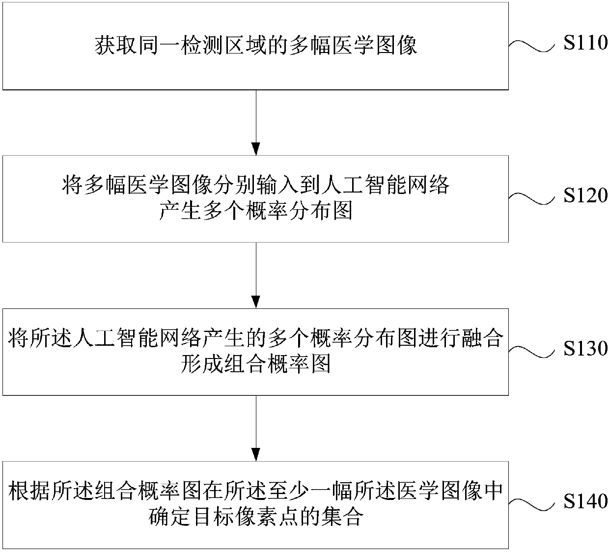
[0045] figure 1 It is a flowchart of a medical image processing method provided by Embodiment 1 of the present invention. The technical solution of this embodiment can be applied to the detection of target pixel points, for example, the target pixel points can be pixels in the lesion area, or can be pixels in any region of interest. The method specifically includes the following operations:

[0046] S110. Acquiring multiple medical images of the same detection area. Optionally, each medical image may contain pixels with different gray values.

[0047] The medical image may be a lung image, and a lung nodule region, an emphysema region, or a rib fracture region can be detected from the medical image through the detection method of the medical image. Medical images can be one-dimensional (1D) data, two-dimensional (2D) images or three-dimensional (3D) images, for example, 1D data can be electrocardiograms collected by electrocardiography; 2D images can be digital X-ray photog...

Embodiment 2

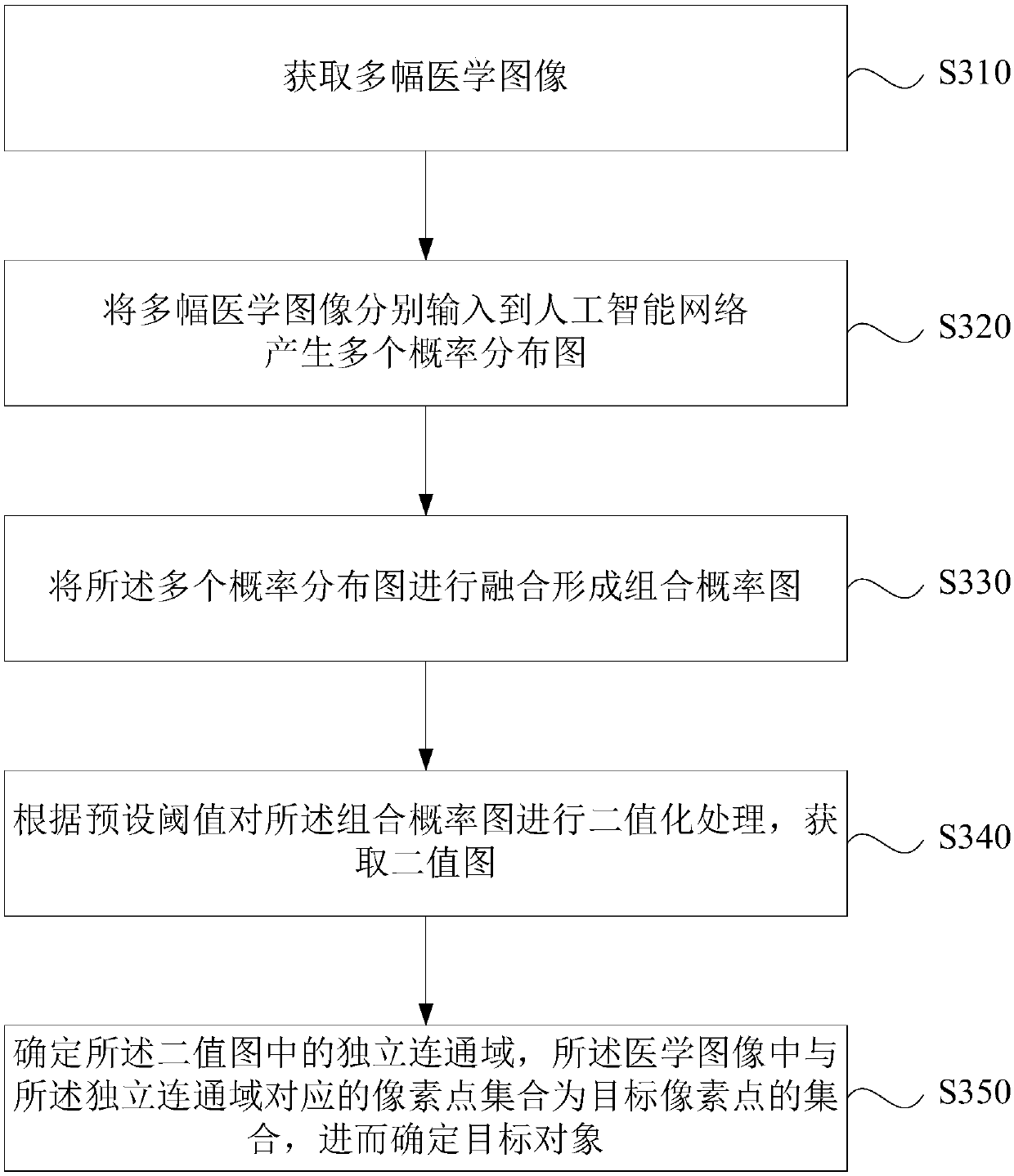
[0063] image 3 It is a flowchart of a medical image processing method provided by Embodiment 2 of the present invention. The technical solution of this embodiment further optimizes the operation of determining the target pixel in at least one of the medical images according to the combination probability map based on any of the above embodiments. Correspondingly, the method of this embodiment includes:

[0064] S310. Acquire multiple medical images, where the multiple medical images correspond to the same target area, and each medical image includes pixels with different gray values. In this embodiment, one of the multiple medical images may be an original image, and the rest of the medical images are original images obtained through image enhancement processing. Further, the enhancement of the medical image is only locally enhanced, and the enhanced medical images have different contrasts.

[0065] S320. Input multiple medical images to the artificial intelligence network...

Embodiment 3

[0075] Figure 4a It is a flowchart of a medical image processing method provided by Embodiment 3 of the present invention. On the basis of any of the above-mentioned embodiments, the technical solution of this embodiment further defines that the multiple medical images are 2D images or slice images, the artificial intelligence network is selected as an end-to-end network, and the multiple medical images are optimized. Operation of Medical Image Input End-to-End Network. Correspondingly, the method of this embodiment includes:

[0076] S410. Acquire multiple medical images, the multiple medical images correspond to the same target area, and each medical image includes multiple slice images, and each slice image includes pixels with different gray values.

[0077] Wherein, the plurality of medical images may include an original medical image and an enhanced image of the original medical image.

[0078] Correspondingly, the method also includes:

[0079] Gaussian filtering i...


PUM


Abstract

The embodiment of the invention discloses a medical image processing method and a medical image processing system. The method includes: a plurality of medical images in a same detection area are obtained; the plurality of medical images are inputted into an artificial intelligence network to generate a plurality of probability distribution maps, wherein the probability distribution maps are used for determining the probabilities of belonging to target pixel points by pixel points in the medical image; carrying out fusion of the plurality of probability distribution maps to form a combined probability map; and determining a set of the target pixel points in at least one of the medical image according to the combined probability map. Therefore, the detection rate and the accuracy of the target pixel point are improved.

Description

technical field [0001] Embodiments of the present invention relate to image processing technologies, and in particular to a medical image processing method and a medical image processing system. Background technique [0002] When performing medical image detection, there are usually two situations in the detection results, one is the real target area, that is, the body position of the actual lesion; the other is a false positive, that is, the body position that does not actually have a lesion but the detection result is a lesion. Therefore, when performing medical image detection, filtering out false positives is crucial for correctly detecting the location of lesions. [0003] Existing methods for lesion detection first use a sliding window for initial detection, and then use a convolutional neural network to remove false positives. Due to the fact that the proportion of lesions and false positives in the image is very small, when the size of the window is too large, there...

Claims


Application Information

Patent Timeline
Patent Type & Authority Applications(China)
IPC IPC(8): G06T7/00G06T7/136G06T7/187G06T7/30G06N3/04
CPCG06T7/0012G06T7/136G06T7/187G06T7/30G06T2207/10081G06T2207/10124G06T2207/30061G06N3/045
Inventor 王季勇李强
Owner SHANGHAI UNITED IMAGING HEALTHCARE
Why Eureka
Try Eureka