Segmentation method and device for blood vessels in fundus image, and storage medium

A fundus image and blood vessel technology, which is applied in the field of image analysis, can solve the problems of large amount of calculation, long processing time and low accuracy, and achieves the effect of good calculation efficiency and improved sensitivity.

Active Publication Date: 2018-06-12
JILIN UNIV
View PDF2 Cites 24 Cited by
  • Summary
  • Abstract
  • Description
  • Claims
  • Application Information

AI Technical Summary

Problems solved by technology

[0006] However, the accuracy of the above method is not high. Although some schemes have high accuracy, the amount of calculation is too large and the processing time is long.

Method used


Image

  • Segmentation method and device for blood vessels in fundus image, and storage medium
  • Segmentation method and device for blood vessels in fundus image, and storage medium
  • Segmentation method and device for blood vessels in fundus image, and storage medium

Examples


Embodiment 1

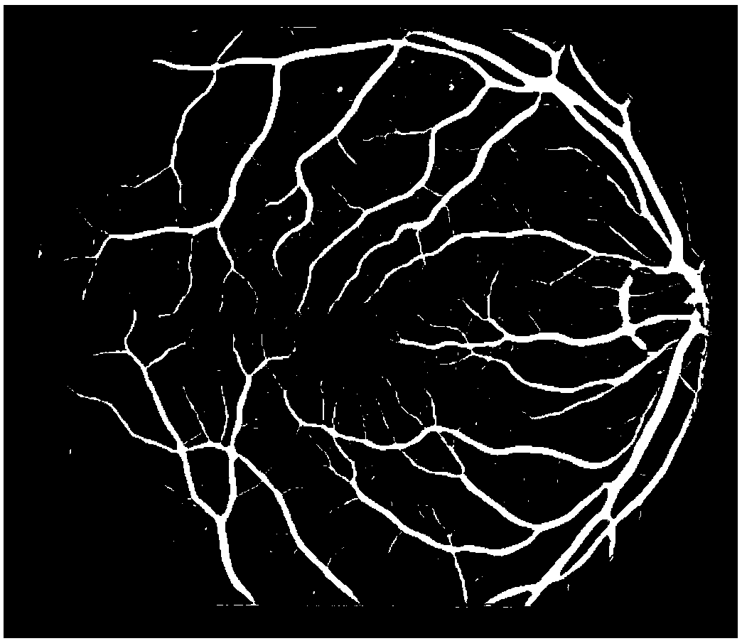
[0103] For blood vessel segmentation in DRIVE and STARE fundus databases, in Figure 11 and Figure 12 The (a) original fundus image, (b) the segmentation result based on the Hessian matrix, (c) the segmentation result based on multi-directional morphology and filtering, (d) the final segmentation result, and (e) are shown in each column. Gold standard chart. From this, it can be seen that the blood vessel segmentation method based on Hessian matrix enhancement in the process of the present invention and the blood vessel segmentation based on multi-directional morphology and filtering have their own advantages and disadvantages. In the present invention, the results of the two methods are merged to finally obtain better results. split effect.

[0104] The segmentation of blood vessels can be regarded as the process of pixel labeling, that is, the process of marking whether the pixels belong to blood vessel points or background points, or called a binary classification proces...


PUM


Abstract

The invention provides a segmentation method and device for blood vessels in a fundus image, and a storage medium. The method comprises the steps that blood vessel segmentation based on Hessian matrixenhancement is conducted, and a threshold value is adopted for segmentation of the blood vessels; a multi-directional linear structure element is used for open operation processing on a G channel, morphological reconstruction is conducted for further enhancement, the multi-directional open operation is adopted and the minimum response is taken to obtain a background without a linear structure, and two images are subtracted to obtain a main vascular network; secondly, multi-directional Gaussian smoothing filtering and multi-directional Gaussian-Laplace filtering are conducted successively, andmulti-directional morphology and morphological reconstruction are adopted to enhance and preserve the filtered vascular network; finally an adaptive threshold value is determined to segment the bloodvessels; the segmentation results of the above two stages are combined to merge and make the final repair to obtain a final blood vessel binary image. The blood vessel segmentation method can improvethe sensitivity of blood vessel recognition and has better calculation efficiency without the need to train in advance.

Description

technical field [0001] The present application relates to the field of image analysis, in particular to a method, device and storage medium for accurately segmenting blood vessels in color fundus images. Background technique [0002] Obtaining blood vessels from fundus images, locating and segmenting blood vessels is the most urgent requirement for fundus image measurement. For the segmentation of blood vessels in the prior art, the conventional method is to segment blood vessels by using image processing methods such as image preprocessing, filtering, fitting, and morphology. The above-mentioned methods have certain effects, but still have certain defects. [0003] For example, multi-scale filtering has a better enhancement effect on blood vessels. Kovács (Kovács G, Hajdu A.Aself-calibrating approach for the segmentation of retinal vessels by templatematching and contour reconstruction[J].Medical image analysis,2016,29:24-46) etc. use matched filtering and contour reconstr...

Claims


Application Information

Patent Timeline
Patent Type & Authority Applications(China)
IPC IPC(8): G06T7/194G06T7/155G06T7/136G06T5/40G06T5/30G06T5/00
CPCG06T5/002G06T5/30G06T5/40G06T7/136G06T7/155G06T7/194G06T2207/30041G06T2207/30101
Inventor 王欣黄卓彦
Owner JILIN UNIV
Why Eureka
Try Eureka