Kit and method for diagnosis of gastric cancer using analysis of bacteria meta-genome

A kit and technology for gastric cancer, applied in the direction of biochemical equipment and methods, microbial determination/testing, etc.

Pending Publication Date: 2018-06-29
MD HEALTHCARE INC
View PDF4 Cites 9 Cited by
  • Summary
  • Abstract
  • Description
  • Claims
  • Application Information

AI Technical Summary

Problems solved by technology

However, regarding the occurrence of gastric cancer, there is no research on the identification of causative factors of gastric cancer from human-derived fluids (such as blood, urine, etc.) method reporting

Method used


Image

  • Kit and method for diagnosis of gastric cancer using analysis of bacteria meta-genome
  • Kit and method for diagnosis of gastric cancer using analysis of bacteria meta-genome
  • Kit and method for diagnosis of gastric cancer using analysis of bacteria meta-genome

Examples


Embodiment 1

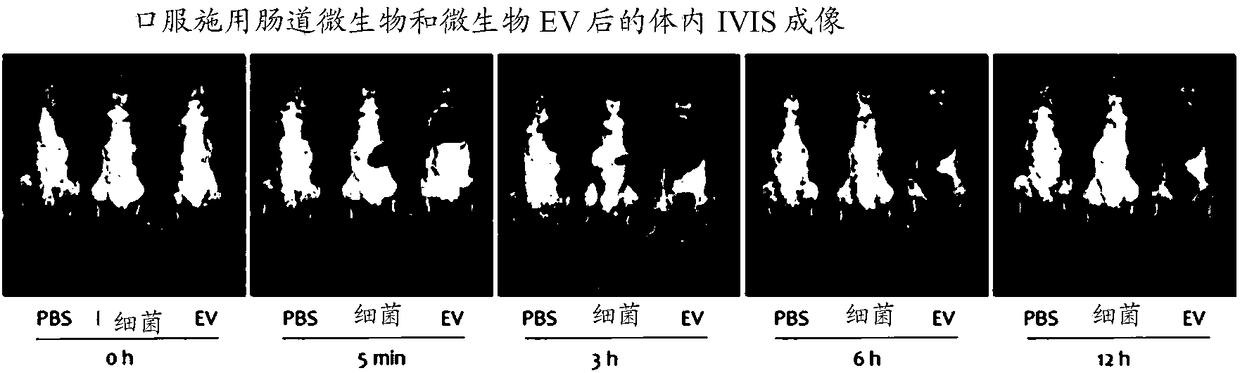
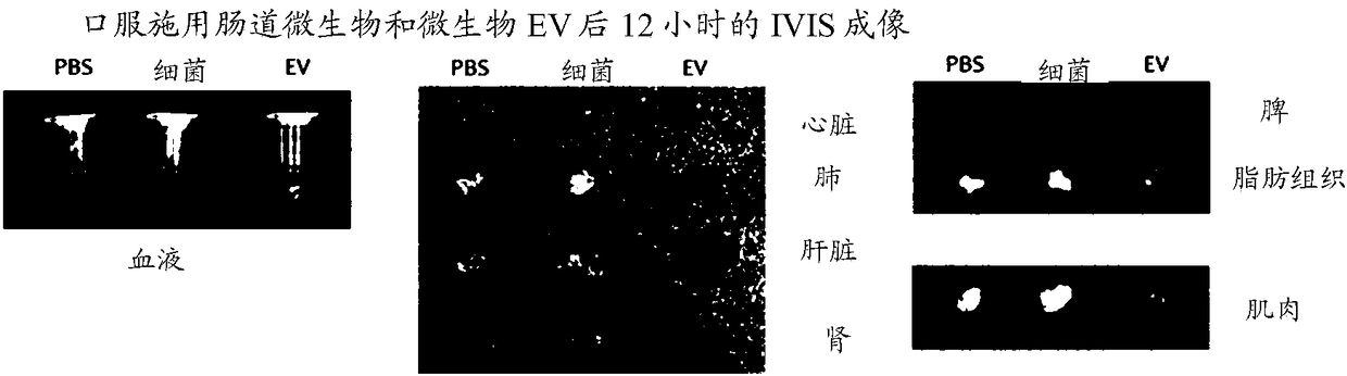
[0083] Example 1. Analysis of In vivo Absorption, Distribution and Excretion Patterns of Enterobacteria and Bacteria-Derived Extracellular Vesicles

[0084] To assess whether enterobacteria and bacterial-derived extracellular vesicles are systemically absorbed through the gastrointestinal tract, experiments were performed using the following method. More specifically, 50 μg each of fluorescently labeled Pseudomonas intestinal bacteria and extracellular vesicles (EVs) derived from the bacteria were orally administered to the gastrointestinal tract of mice, and at 0 hours, and at Fluorescence was measured after 5 minutes, 3 hours, 6 hours and 12 hours.

[0085] As a result of observing the overall image of the mouse, such as Figure 1A As shown, bacteria were not absorbed systemically at the time of administration, but EVs derived from bacteria were absorbed systemically at 5 minutes after administration, and strong fluorescence was observed in the bladder at 3 hours after admin...

Embodiment 2

[0087] Example 2. Vesicle Isolation and DNA Extraction from Blood

[0088] To isolate vesicles and extract DNA from blood and urine, first add blood and urine to a 10 ml tube, centrifuge at 3,500 x g and 4 °C for 10 min, pellet the suspension, and collect only the supernatant , and place it in a new 10ml tube. The collected supernatant was filtered with a 0.22 μm filter to remove bacteria and impurities, then placed in a central centrifugal filter (50 kD) and centrifuged at 1500 × g and 4° C. for 15 minutes to discard material with a size less than 50 kD, Then concentrate to 10ml. Bacteria and impurities were removed again using a 0.22 μm filter, and then the resulting concentrate was subjected to ultracentrifugation at 150,000 × g and 4° C. for 3 hours to remove the supernatant, and the aggregated precipitate was washed with phosphate buffer ( PBS) to obtain vesicles.

[0089] 100 μl of vesicles isolated from serum according to the method described above were boiled at 100...

Embodiment 3

[0092] Example 3. Bacterial metagenomic analysis using DNA from vesicles extracted from blood

[0093] As shown in Table 2 below, using the same method as in Example 2, using blood samples from 67 patients with gastric cancer and 198 normal individuals, urine samples from 61 patients with gastric cancer and 120 normal individuals, the gender and Age-matched, they were then subjected to PCR using 16S rDNA primers to amplify the DNA and subsequently sequenced (Illumina MiSeq sequencer). Output the results as a Standard Flow Diagram (SFF) file, and convert the SFF file to a sequence file (.fasta) and nucleotide quality score file using GS FLX software (v2.9), then determine credit ratings for reads, And fractions with window (29 bps) average base call accuracy less than 99% (Phred score < 20) were removed. After removal of low-quality parts, only reads with a length of 300 bps or greater (Sickle version 1.33) were used, and for operational taxonomic unit (OTU) analysis clusterin...


PUM


Abstract

The present invention relates to a kit and method for diagnosis of gastric cancer using analysis of bacteria meta-genome. Provided are a kit and method for providing information on the diagnosis of gastric cancer through a bacterial meta-genome analysis. More specifically, the bacterial meta-genome analysis is executed on the genomes present in the vesicles isolated from a sample derived from an inspection target to analyze the increase or decrease in the content of the extracellular vesicles derived from specific bacteria, thereby diagnosing gastric cancer and predicting the risk of the gastric cancer. According to the present invention, the meta-genomes of EVs derived from bacteria present in the human-derived sample are analyzed to predict the risk of gastric cancer, thereby predictingand diagnosing the risk groups of gastric cancer to prevent or delay the incidence of gastric cancer through appropriate management. The present invention also enables early prediction even after theincidence to lower the incidence rate of gastric cancer and enhance the treatment effects.

Description

[0001] Cross References to Related Applications [0002] This application claims the priority and benefit of Korean Patent Application No. 2016-0172584 filed on December 16, 2016 and Korean Patent Application No. 2017-0072432 filed on June 9, 2017, the disclosures of which are incorporated by reference in their entirety into this article. technical field [0003] The present disclosure relates to kits and methods for providing information for the diagnosis of gastric cancer through bacterial metagenomic analysis, and more particularly, to a method for analyzing bacterial metagenomic analysis using a sample derived from a subject, by analyzing specific A kit and method for predicting the risk of gastric cancer or diagnosing gastric cancer by increasing or decreasing the content of extracellular vesicles of sexual bacteria. Background technique [0004] Globally, the incidence of gastric cancer is high in East Asia including South Korea, China, and Japan, while it is relative...

Claims


Application Information

Patent Timeline
Patent Type & Authority Applications(China)
IPC IPC(8): C12Q1/6869C12Q1/6886
CPCC12Q1/6886C12Q2600/158C12Q1/6869C12Q1/689C12Q2531/113C12Q1/6895
Inventor 金润根全成圭
Owner MD HEALTHCARE INC
Why Eureka
Try Eureka