Ultrasonic cardiac assessment of hearts with medial axis curvature and transverse eccentricity

A curved, heart-shaped technology, applied in the field of medical diagnostic ultrasound systems, which can solve problems such as irregular spacing of editing controls and difficulties in drawing boundaries

Active Publication Date: 2018-07-17
KONINKLJIJKE PHILIPS NV
View PDF20 Cites 0 Cited by
  • Summary
  • Abstract
  • Description
  • Claims
  • Application Information

AI Technical Summary

Problems solved by technology

Lateral eccentricity of the heart can make boundary drawing more difficult and editing controls more irregularly spaced

Method used


Image

  • Ultrasonic cardiac assessment of hearts with medial axis curvature and transverse eccentricity
  • Ultrasonic cardiac assessment of hearts with medial axis curvature and transverse eccentricity
  • Ultrasonic cardiac assessment of hearts with medial axis curvature and transverse eccentricity

Examples


Embodiment Construction

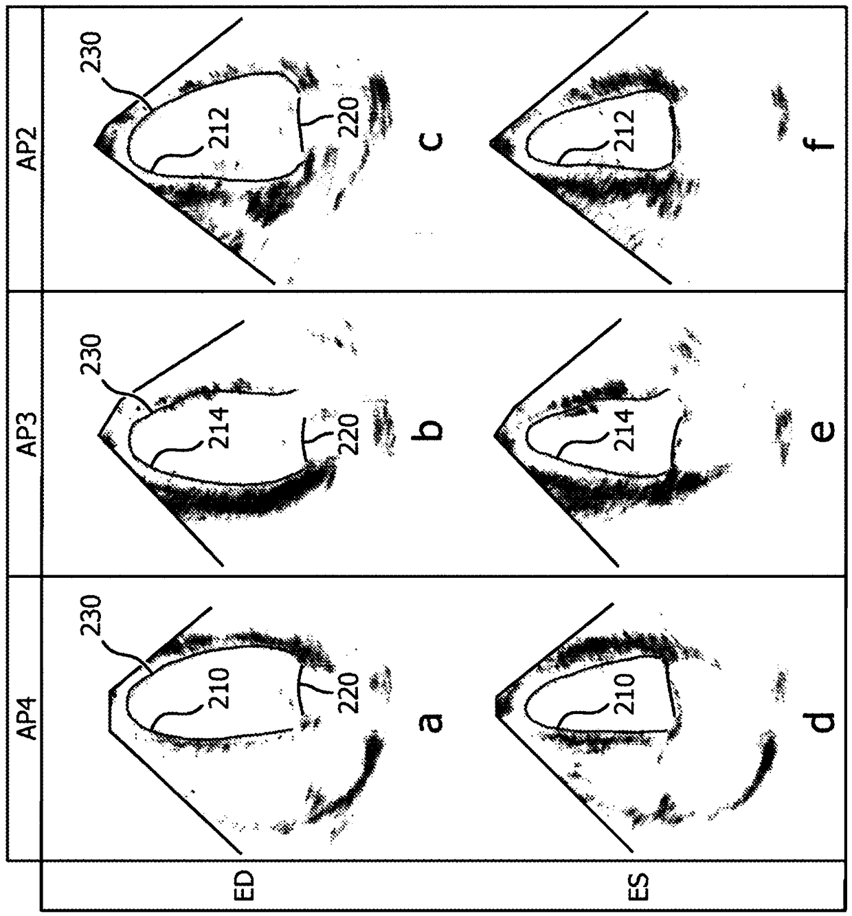
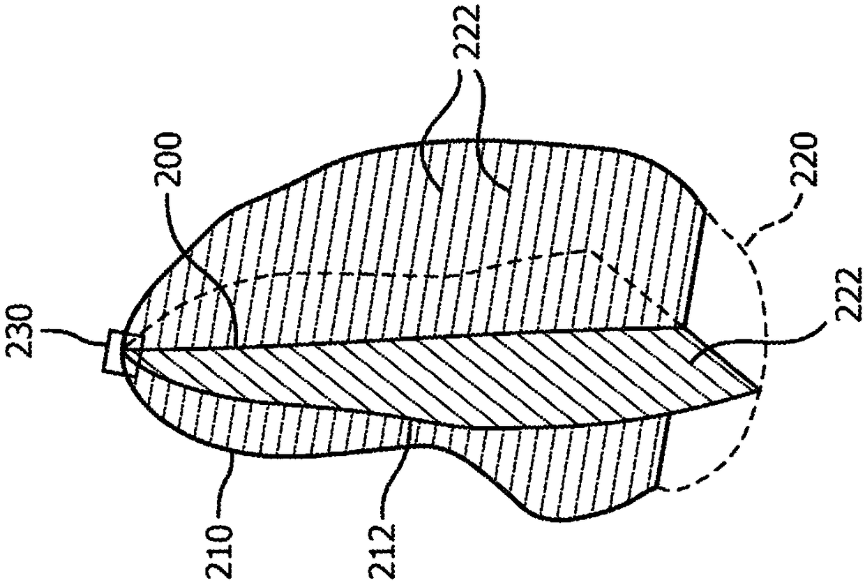
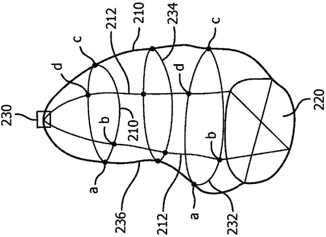
[0019] In accordance with the principles of the present invention, an ultrasound diagnostic system and method are described that correct for cardiac curvature and lateral decentration in ultrasound cardiac images. The generally straight median axis is drawn as a curve spaced evenly between the chamber walls and extending from the apex to the plane of the mitral valve. Reduce lateral decentering by stretching the myocardium in the image to produce a more uniform shape. As a result of these measures, the boundaries of the heart chambers are rendered such that the chamber walls are more equidistant and orthogonal to the viewing plane, enabling the creation of a more complete long-axis MPR view and allowing in-plane editing of the transverse MPR plane to represent segmentation boundaries and More isometric and orthogonal displacement of control points. An unshortened long-axis MPR view is generated that is a curved slice through the curved medial axis. Editing control points in ...


PUM


Abstract

An ultrasonic imaging system produces more diagnostic cardiac images of the left ventricle by plotting the longitudinal medial axis of the chamber between the apex and mitral valve plane as a curved line evenly spaced between the opposite walls of the myocardium. Transverse image planes are positioned orthogonal to the curved medial axis with control points positioned in the short axis view on lines evenly spaced around and emanating from the medial axis. If the short axis view is of an oval shaped chamber the transverse image is stretched to give the heart a more rounded appearance resultingin better positioning of editing control points.

Description

technical field [0001] The present invention relates to medical diagnostic ultrasound systems, and in particular to ultrasound systems for performing cardiac evaluation on hearts with shaft curvature and transverse irregularities. Background technique [0002] Ultrasound imaging is widely used to evaluate and diagnose the condition of the heart. An ultrasound probe is pressed against the patient's body just below the chest cavity and aimed upwards to view the heart from the apex. A sequence of three-dimensional image datasets is acquired over a cardiac cycle while the heart is fully in view of the live image. The three-dimensional image data is analyzed by a segmentation processor to identify various features and properties of the heart, such as the endothelial border of the left ventricle, ejection fraction, and myocardial motion. Two such processors for cardiac analysis are the automatic boundary detection processors described in US Pat. No. 6,491,636 (Chenal et al.) and...

Claims


Application Information

Patent Timeline
Patent Type & Authority Applications(China)
IPC IPC(8): A61B8/08A61B8/00G06T7/00
CPCA61B8/0883A61B8/466A61B8/467A61B8/483A61B8/52A61B8/523A61B8/5269G06T19/00G06T2207/10136G06T2207/20096G06T2207/30048G06T2207/30172G06T2210/41G06T2219/008G06T7/12G06T7/149A61B8/463A61B8/5253G06T13/20G06T19/20G06T2219/2021
Inventor S·H·塞特尔迈尔D·普拉特R·J·施耐德
Owner KONINKLJIJKE PHILIPS NV
Why Eureka
Try Eureka