
Segmentation method of GA lesion in sd-oct image based on deep voting model

A SD-OCT and model technology, applied in the field of lesion segmentation, can solve the problem of difficult to obtain ideal results and affect the accuracy of lesion segmentation, and achieve the effect of breaking through the bottleneck of segmentation dependence, breaking through sensitivity, and improving segmentation accuracy.

Active Publication Date: 2021-12-28
NANJING UNIV OF SCI & TECH
View PDF3 Cites 0 Cited by
  • Summary
  • Abstract
  • Description
  • Claims
  • Application Information

AI Technical Summary

Problems solved by technology

However, since retinal diseases usually change the structure of retinal tissue layers, it is difficult for existing layer segmentation methods to obtain ideal results, which in turn affects the final lesion segmentation accuracy

Method used


Image

  • Segmentation method of GA lesion in sd-oct image based on deep voting model
  • Segmentation method of GA lesion in sd-oct image based on deep voting model
  • Segmentation method of GA lesion in sd-oct image based on deep voting model

Examples


Embodiment Construction

[0021] The present invention will be further described below in conjunction with the accompanying drawings.

[0022] combine figure 1 , the SD-OCT retinal image GA lesion segmentation method based on depth voting model of the present invention comprises the following steps:

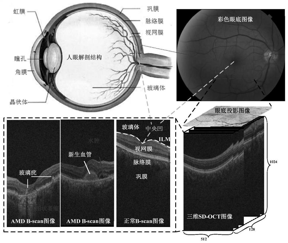
[0023] Step 1. Collect SD-OCT retinal images, and use existing OCT imaging equipment to collect retinal images. The imaging area of ​​the SD-OCT image, the comparison with the color fundus image, the imaging results, and the manifestations of the lesions such as figure 2 shown.

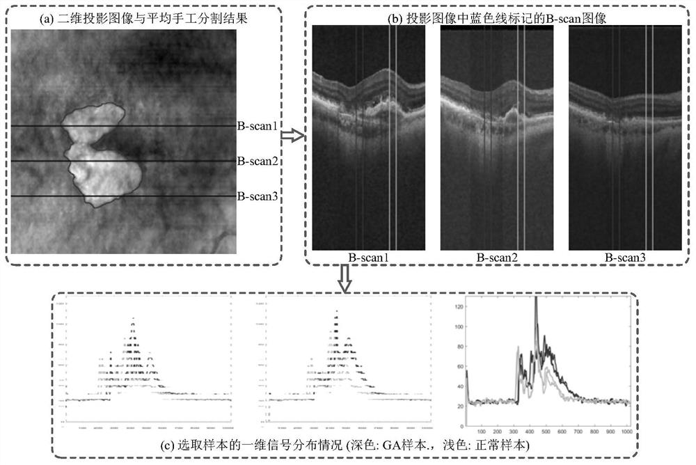
[0024] Step 2. Obtain labeled samples according to the standard data set of GA lesions.

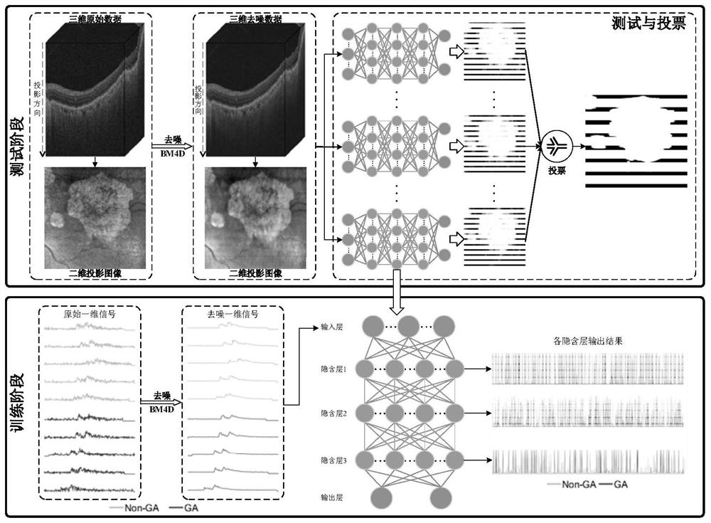
[0025] Step 3, use the BM4D algorithm to perform denoising processing on the original 3D data. The denoising results of 3D, 2D and 1D signals are as follows: figure 1 shown.

[0026] Step 4. On the basis of the denoising data, randomly extract positive and negative labeled samples from the labeled sample set to construct a training da...


PUM


Abstract

The invention discloses a SD-OCT image GA lesion segmentation method based on a deep voting model. The method first constructs a five-layer deep network model, the first layer is the input layer, the fifth layer is the output layer, and the hidden layer is composed of three layers of sparse auto-encoders. In the training phase, the labeled positive and negative samples with 1024-dimensional features are directly input into the deep network model, and the deep description features of the data are captured by an unsupervised three-layer autoencoder, and obtained by training the soft-max classifier of the output layer. the final segmentation result. Finally, a voting decision strategy is used to improve the segmentation results of the ten trained models. By using a deep network model to express the complex data structure in 3D data, this method breaks through the bottleneck of traditional methods for image layer segmentation, and breaks through the sensitivity of traditional methods to data from different sources, and achieves high-precision quantitative analysis of GA lesions It has important practical significance for the prevention and diagnosis of age-related macular degeneration.

Description

technical field [0001] The invention relates to a lesion segmentation method, in particular to a frequency domain optical coherence tomography retinal image geographic atrophy lesion segmentation method based on a depth voting model. Background technique [0002] Among retinal diseases, age-related macular degeneration (Age-related Macular Degeneration, AMD) has become one of the main diseases affecting the health and quality of life of the elderly. SD-OCT retinal image is a frequency-domain optical coherence tomography image, which can effectively present changes in the gray scale and structure of retinal tissue layers. Clinical experiments have shown that SD-OCT retinal images can show more and more AMD Retinopathy caused by tissue, such as drusen, geographic atrophy, edema, retinal detachment, etc. Due to the unique layered structure of SD-OCT retinal images, it is difficult to obtain ideal analysis results by traditional methods. Since GA is often associated with retin...

Claims


Application Information

Patent Timeline
Patent Type & Authority Patents(China)
IPC IPC(8): G06T7/00G06T7/11G06K9/62
CPCG06T7/0012G06T7/11G06T2207/30041G06T2207/20081G06T2207/10101G06F18/241
Inventor 纪则轩陈强
Owner NANJING UNIV OF SCI & TECH
Learn More