Method and system for choroidal vessel segmentation based on three-dimensional coherence tomography images

A technology of coherence tomography and tomographic images, applied in the field of quantitative analysis of three-dimensional medical images, can solve the problems of inability to accurately segment three-dimensional choroidal blood vessels, inability to analyze choroidal structure, etc., to improve work accuracy and efficiency, high segmentation accuracy, and repeatability rate-optimized effect

Active Publication Date: 2022-06-21
武汉华悦立远科技有限公司
View PDF5 Cites 0 Cited by
  • Summary
  • Abstract
  • Description
  • Claims
  • Application Information

AI Technical Summary

Problems solved by technology

[0004] Existing quantitative analysis methods are either unable to analyze the choroidal structure, or can only perform incomplete analysis on the two-dimensional OCT interface, and they cannot accurately segment the three-dimensional choroidal vessels to calculate various clinical indicators based on the volume of the choroidal vessels

Method used


Image

  • Method and system for choroidal vessel segmentation based on three-dimensional coherence tomography images
  • Method and system for choroidal vessel segmentation based on three-dimensional coherence tomography images
  • Method and system for choroidal vessel segmentation based on three-dimensional coherence tomography images

Examples


Embodiment Construction

[0027] In order to make the objectives, technical solutions and advantages of the present invention more clearly understood, the present invention will be described in further detail below with reference to the accompanying drawings and embodiments. It should be understood that the specific embodiments described herein are only used to explain the present invention, not for The invention is limited.

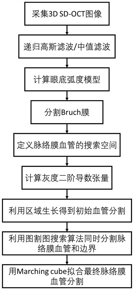
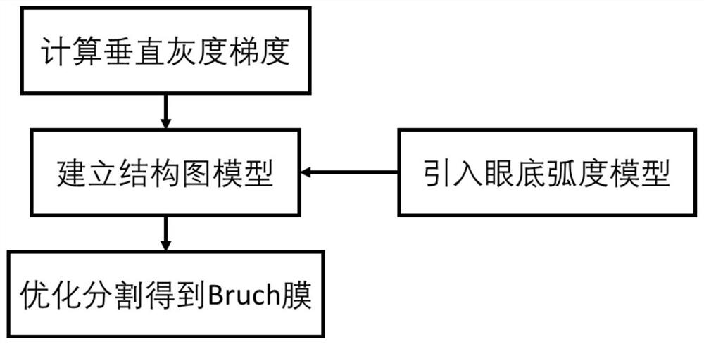
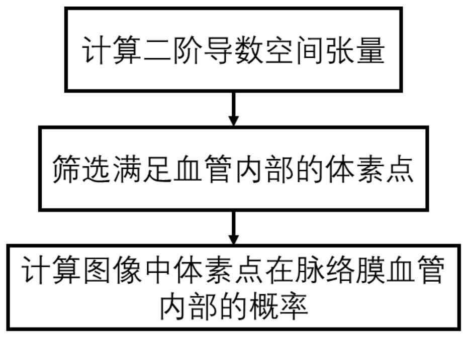
[0028] like figure 1 As shown, the present invention provides a choroidal blood vessel segmentation method based on three-dimensional coherence tomography images. Specifically, the choroidal blood vessel segmentation method based on three-dimensional coherence tomography images includes the following steps:

[0029] S1. Collect a three-dimensional frequency-domain coherent optical tomographic image, and use recursive Gaussian filtering and median filtering to eliminate noise information in the three-dimensional coherent tomographic image.

[0030] First, collect three-dimensiona...


PUM


Abstract

The invention discloses a choroidal vessel segmentation method and system based on a three-dimensional coherent tomography image, which collects a three-dimensional frequency-domain coherent optical tomographic image; The fundus arc model is calculated from the actual size; the retinal base layer Bruch's membrane is segmented from the fundus arc model using a single-layer graph search method; the spatial tensor of each voxel point is calculated to obtain the point probability of the voxel point inside the choroidal vessel , screen out the pre-selected area of ​​the choroidal vessel with a point probability value greater than 0; screen out the point probability value greater than 75% and the point probability value less than 25% from the pre-selected area of ​​the choroidal vessel as the high and low thresholds for the growth of the pre-selected area, and then obtain the choroid Initial segmentation of foreground and background of blood vessels; according to the initial segmentation of foreground and background of choroidal vessels, the optimal model of the structure graph is solved to obtain accurate segmentation of choroidal vessels.

Description

technical field [0001] The invention relates to a three-dimensional medical image quantitative analysis technology, in particular to a choroidal blood vessel segmentation method and system based on three-dimensional coherence tomography images. Background technique [0002] Spectral-Domain Optical Coherence Tomography (SD-OCT) is an imaging technology that can produce three-dimensional micron-level precision images. Compared with traditional high-energy radiation (X-ray, CT, etc.), OCT provides non-invasive, harmless scanning imaging with low-energy near-infrared light. It is the only technology that can realize the reconstruction of living 3D human eye images at this stage. During the imaging process, the detection light has to pass through the optic nerve fiber layer and the retinal pigment epithelial cell layer with high scattering before reaching the choroid, so it is difficult to image the choroid with OCT. In recent years, there have been only a few studies on the ch...

Claims


Application Information

Patent Timeline
Patent Type & Authority Patents(China)
IPC IPC(8): G06T7/194G06T5/00G06T7/136
CPCG06T5/002G06T7/136G06T7/194G06T2207/30101G06T2207/10076G06T2207/10081G06T2207/20032
Inventor 张立付峥
Owner 武汉华悦立远科技有限公司
Why Eureka
Try Eureka