Multimodal nuclear magnetic resonance image segmentation method for glioblastoma

A technology for glioblastoma and nuclear magnetic resonance images, which is applied in the field of digital medical image analysis and intelligent health management, can solve problems such as subdivision, and achieve the effect of reducing data acquisition and storage

Active Publication Date: 2018-08-24
ZHEJIANG CHINESE MEDICAL UNIVERSITY
View PDF1 Cites 18 Cited by
  • Summary
  • Abstract
  • Description
  • Claims
  • Application Information

AI Technical Summary

Problems solved by technology

However, through the analysis of the existing GBM multimodal magnetic resonance image segmentation methods, it is found that although the above studies have achieved certain research results, they all focus on searching and segmenting the entire tumor area, and do not further subdivide the entire tumor into tumors. sub-region

Method used


Image

  • Multimodal nuclear magnetic resonance image segmentation method for glioblastoma
  • Multimodal nuclear magnetic resonance image segmentation method for glioblastoma
  • Multimodal nuclear magnetic resonance image segmentation method for glioblastoma

Examples


Embodiment 1

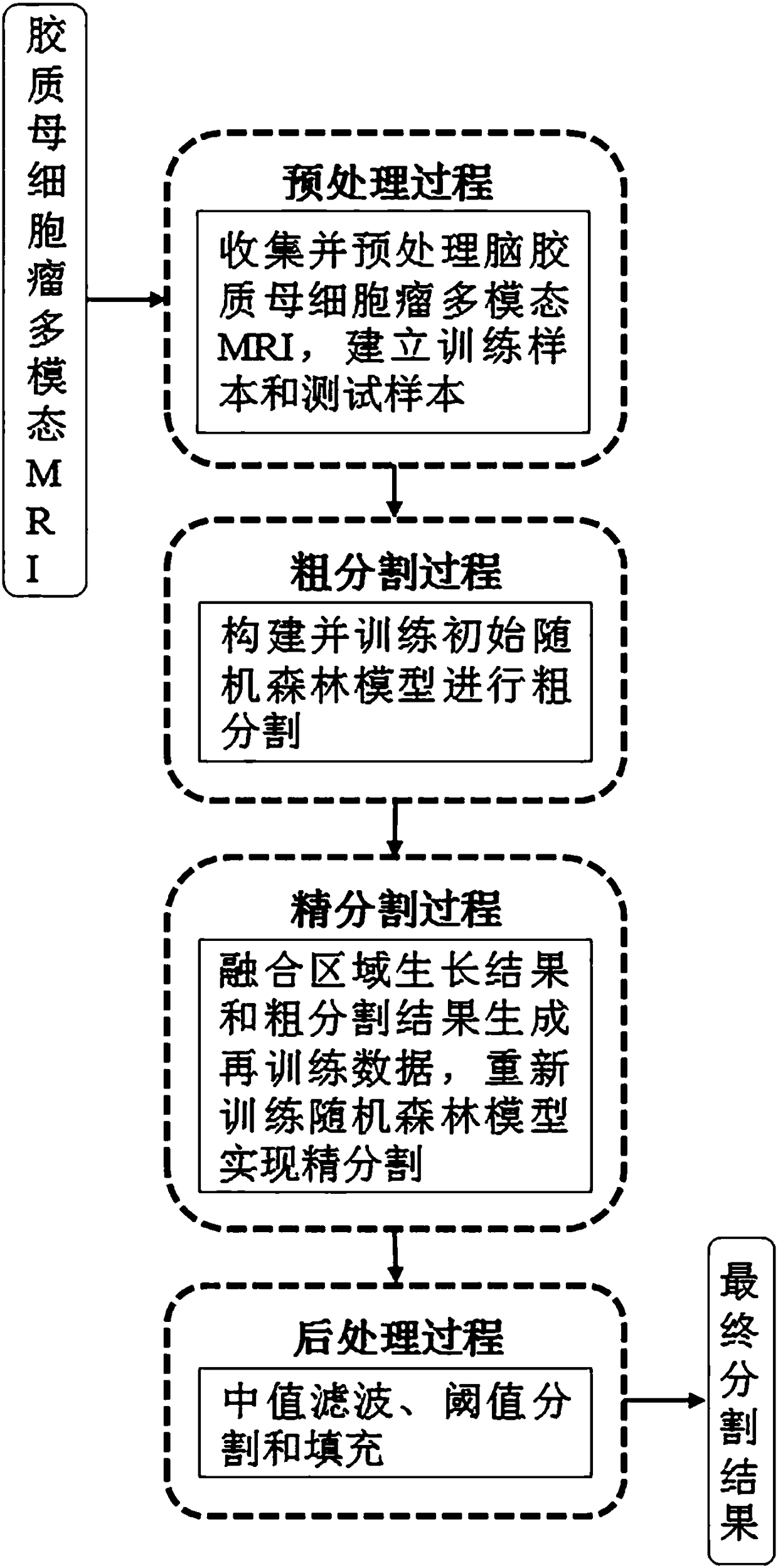
[0074] Embodiment 1, the multimodal nuclear magnetic resonance image segmentation method of brain glioblastoma, such as Figure 1-6 As shown, the multimodal nuclear magnetic resonance image (hereinafter referred to as MRI for short) includes three modal image information of T1W image before contrast agent injection (T1), T1W image after contrast agent injection (T1c), and FLAIR image.

[0075] According to the method of random forest and region growing, the present invention divides the MRI image of the brain into normal tissue area, necrosis area, active tumor area, T1 abnormal area (excluding necrosis area and active tumor area) and FLAIR abnormal area (excluding Including necrosis area, active tumor area and T1 abnormal area) 5 parts, there is no intersection area in the above 5 segmentation areas. Random forest has the characteristics of few parameters that need to be adjusted, high computing speed, strong anti-noise ability and no over-fitting phenomenon.

[0076] Since ...


PUM


Abstract

The invention provides a multimodal nuclear magnetic resonance image segmentation method for glioblastoma. The segmentation strategy combining the random forest method and the regional growth method is employed, the result of regional growth segmentation of a glioma multimodal magnetic resonance image is replaced with the corresponding random forest segmentation result with low confidence, retraining data is generated to re-train a random forest model, fine segmentation of the glioma multimodal magnetic resonance image is carried out, and a brain MRI image is segmented into a normal brain tissue area, a necrotic area, an active tumor area, a T1 abnormal area and a FLAIR abnormal area. The method is advantaged in that through fine segmentation and positioning of the glioblastoma, doctors are assisted in diagnosis and other treatment tasks, accurate positioning of the glioblastoma and more accurate fine segmentation of different tumor sub areas are carried out, the doctors are facilitated to diagnose the glioblastoma more quickly and accurately, and an accurate treatment scheme is made.

Description

technical field [0001] The invention relates to the fields of digital medical image analysis and intelligent health management, in particular to a multimodal nuclear magnetic resonance image segmentation method for glioblastoma. Background technique [0002] Glioma is the most common primary brain tumor, more than half of which are the most malignant glioblastoma (Glioblastoma Multiforme, GBM). For GBM, no matter its form is benign or malignant, it will increase intracranial pressure, compress brain tissue, cause damage to the central nervous system, and endanger the life of the patient. The location and quantitative calculation of GBM lesion tissue (such as tumor volume, etc.) are crucial for the diagnosis, surgical planning, and postoperative analysis of GBM. In clinical practice, radiologists usually manually segment tumors from multimodal magnetic resonance images, which is tedious and time-consuming. Automatically segmenting GBM tumors can not only free doctors from i...

Claims


Application Information

Patent Timeline
Patent Type & Authority Applications(China)
IPC IPC(8): G06T7/11G06T7/13G06N3/08G06N3/04
CPCG06N3/08G06T7/11G06T7/13G06T2207/10088G06T2207/20032G06T2207/30016G06T2207/30096G06N3/045
Inventor 赖小波高卫红李文胜黄燕吕莉莉
Owner ZHEJIANG CHINESE MEDICAL UNIVERSITY
Why Eureka
Try Eureka