Method, device, equipment for segmentation of lesion in biological image and storage medium

A technology of image center and focus, applied in the field of image processing, can solve the problems of missed diagnosis, misdiagnosis, strong subjectivity, time-consuming and laborious, etc.

Active Publication Date: 2018-10-19
讯飞医疗科技股份有限公司
View PDF8 Cites 44 Cited by
  • Summary
  • Abstract
  • Description
  • Claims
  • Application Information

AI Technical Summary

Problems solved by technology

[0003] In view of this, the present invention provides a lesion segmentation method, device, equipment and storage medium in a biological image, which are used to solve the time-consuming and labor-intensive method of locating a lesion in a biological image in the prior art, and are highly subjective. , it is easy to cause missed diagnosis and misdiagnosis. The technical solution is as follows:

Method used


Image

  • Method, device, equipment for segmentation of lesion in biological image and storage medium
  • Method, device, equipment for segmentation of lesion in biological image and storage medium
  • Method, device, equipment for segmentation of lesion in biological image and storage medium

Examples


Embodiment Construction

[0082] The following will clearly and completely describe the technical solutions in the embodiments of the present invention with reference to the accompanying drawings in the embodiments of the present invention. Obviously, the described embodiments are only some, not all, embodiments of the present invention. Based on the embodiments of the present invention, all other embodiments obtained by persons of ordinary skill in the art without making creative efforts belong to the protection scope of the present invention.

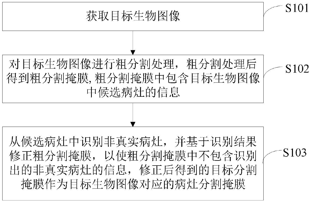
[0083] In view of the fact that the method of manually locating lesions in the prior art is not only time-consuming and laborious, but also highly subjective, which can easily lead to missed diagnosis and misdiagnosis, the embodiment of the present invention provides a method for segmenting lesions in biological images, which can automatically locate lesions , see figure 1 , showing a schematic flow chart of the method, which may include:

[0084] Step S101: ...


PUM


Abstract

The present invention provides a method, device, equipment for the segmentation of a lesion in a biological image and a storage medium. The method comprises a step of acquiring a target biological image, a step of performing coarse segmentation processing on the target biological image and obtaining a coarse segmentation mask after the rough segmentation processing, wherein the coarse segmentationmask includes information of candidate lesions in the target biological image, a step of identifying a non-real lesion from the candidate lesions, correcting the rough segmentation mask based on a recognition result such that the information of an identified non-real lesion is not included in the coarse segmentation mask and a target segmentation mask obtained after the correction is used as a lesion segmentation mask corresponding to the target biological image. According to the method, the device, the equipment and the storage medium, the lesion can be automatically positioned from the target biological image, the mode is labor-saving, the time consumption of the positioning of the lesion is reduced, misdiagnosis and missed diagnosis caused by the manual positioning of the lesion are avoided, the positioned lesion can also assist the doctor to carry out fast and accurate analysis, and the diagnostic efficiency and diagnostic accuracy of doctors are improved.

Description

technical field [0001] The present invention relates to the technical field of image processing, in particular to a lesion segmentation method, device, equipment and storage medium in biological images. Background technique [0002] In the actual clinical diagnosis in the medical field, it is necessary to locate the lesion from the biological image, so as to locate the cause of the lesion. The diagnosis method in the prior art is manual diagnosis, that is, the doctor needs to locate the lesions in the biological image with the naked eye. However, some biological images are very large, but the lesions in the biological image are small, such as fundus images, and the resolution of fundus images The resolution is generally 4000*3000 pixels, and some small lesions in the fundus image may only occupy dozens of pixels, and the size of different lesions is also different, which undoubtedly brings great difficulties to doctors to locate lesions with the naked eye, especially when fa...

Claims


Application Information

Patent Timeline
Patent Type & Authority Applications(China)
IPC IPC(8): G06T7/10G06T7/70
CPCG06T2207/30041G06T7/10G06T7/70
Inventor 王凤艳殷保才刘聪
Owner 讯飞医疗科技股份有限公司
Why Eureka
Try Eureka