Method for detecting cyclic forms of cancer cells

A cell cycle and cancer cell technology, applied in the direction of tumor/cancer cells, animal cells, vertebrate cells, etc., can solve problems such as the inability to recognize cancer cells in essence

Inactive Publication Date: 2018-10-30
SANMING UNIV
View PDF2 Cites 0 Cited by
  • Summary
  • Abstract
  • Description
  • Claims
  • Application Information

AI Technical Summary

Problems solved by technology

[0004] The present invention provides a method for detecting the cycle morphology of cancer cells, aiming at improving the problem of inability to recognize cancer cells in essence in the prior art

Method used


Image

  • Method for detecting cyclic forms of cancer cells
  • Method for detecting cyclic forms of cancer cells

Examples


Embodiment 1

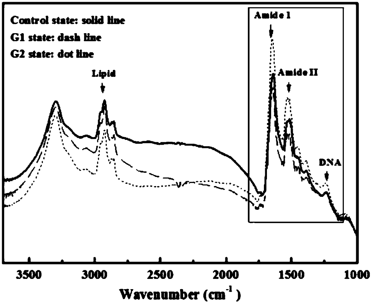
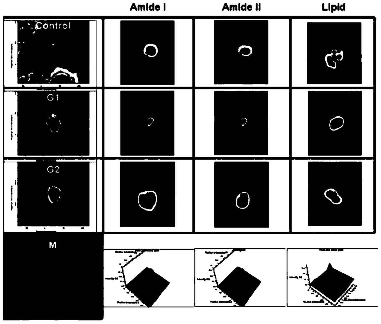
[0032] Embodiment 1, the first embodiment of the present invention provides a method for detecting the cycle morphology of cancer cells. The method for detecting the cycle morphology of cancer cells in this embodiment is based on synchrotron radiation infrared (synchrotron radiation infrared, SR-IR) microscopy Realized by spectral and SR-IR spectral imaging technology, specifically including steps S01 to S03:

[0033] Step S01, extracting four parts of the culture solution containing a certain number of cancer cells from the culture medium, and putting them in different culture bottles, adding a cell cycle blocker to three of the culture bottles and treating them for 24 hours to Make the three cancer cells stay in G1 phase, G2 phase and M phase respectively.

[0034] In a specific implementation, the medium may be RPMI-1640 (Roswell Park Memorial Institute-1640) medium containing 10% heat-inactivated fetal bovine serum and 1% penicillin-streptomycin-L-glutamine, And the mediu...

Embodiment 2

[0047] Embodiment 2, the second embodiment of the present invention provides a method for detecting the cycle morphology of cancer cells, including step S11 to step S16:

[0048] Step S11, extract four culture solutions containing a certain number of cancer cells from the culture medium, and put them in different culture flasks, respectively use 1x phosphate buffered saline with pH 7.4 to culture the four culture flasks. solution for washing and adding fresh culture medium (without cancer cells).

[0049] In specific implementation, a hemocytometer can be used to measure the number of cancer cells in the culture medium.

[0050] Wherein, the medium is RPMI-1640 medium containing 10% heat-inactivated fetal bovine serum and 1% penicillin-streptomycin-L-glutamine, and the medium is at 37°C and contains 5% CO 2 Stored in a humid atmosphere.

[0051] It should be noted that the purpose of washing the medium with 1x phosphate-buffered saline (1xPBS) pH 7.4 is to release cells fro...


PUM


Abstract

The invention provides a method for detecting cyclic forms of cancer cells, and relates to the technical field of biology. The method includes: extracting four parts of nutrient solutions, containingcertain numbers of the cancer cells, from a culture medium, placing the four parts of nutrient solutions into different culture bottles, adding cell cycle retarding agents into three of the culture bottles, and processing for 24 hours to enable the three parts of cancer cells to stay in a G1 phase, a G2 phase and an M phase respectively; slicing a plurality of infrared lantern slides into the sizeof 1cmx1cm, placing the infrared lantern slides, which are disinfected and arranged in an array, into a sterile culture dish, and stacking a glass slide on every infrared lantern slide; dropping thenutrient solutions, in certain cell concentrations, of the four culture bottoms onto different glass slides, subjecting the nutrient solutions on the glass slides to synchrotron radiation infrared microscopic spectral imaging so as to obtain functional group concentration distributions of the four parts of cancer cells and different growing conditions of the cancer cells, and finally realizing substantial acknowledging of the cancer cells.

Description

technical field [0001] The invention relates to the biological field, in particular to a method for detecting the cycle morphology of cancer cells. Background technique [0002] Currently, cancer cells are roughly classified into four types: squamous cell carcinoma, adenocarcinoma, undifferentiated carcinoma, and carcinoma that cannot be classified. Further subdivisions are highly differentiated and lowly differentiated. This kind of distinction is very rough and does not distinguish the essence of cancer cells, but only the appearance of cancer cells with different shapes and changes seen under the microscope. [0003] However, the morphology of cancer cells is different in each cell cycle. Such classification is obviously not enough for understanding and treating cancer cells, which is also the bottleneck of cancer treatment. Therefore, it is urgent to design a set of methods that can detect the cycle morphology of cancer cells in order to recognize cancer cells in essen...

Claims


Application Information

Patent Timeline
Patent Type & Authority Applications(China)
IPC IPC(8): C12N5/09C12Q1/02
CPCC12N5/0693G01N33/5005
Inventor 郑皓远蔡雪霁付红琴林晓哲彭小凤刘希华邢建宏王春宝李奇勇张君诚
Owner SANMING UNIV
Why Eureka
Try Eureka