Proximal femur segmentation method and device, computer device and storage medium

A technology of proximal femur and computer program, which is applied in the computer field to reduce diagnostic interference information, make up for low diagnostic accuracy, and improve diagnostic efficiency

Pending Publication Date: 2018-11-06
PING AN TECH (SHENZHEN) CO LTD
View PDF3 Cites 23 Cited by
  • Summary
  • Abstract
  • Description
  • Claims
  • Application Information

AI Technical Summary

Problems solved by technology

[0003] The main purpose of this application is to provide a method for segmenting the proximal femur, aiming to solve the technical problem of accurately separating the proximal femur in 3D MRI images

Method used


Image

  • Proximal femur segmentation method and device, computer device and storage medium
  • Proximal femur segmentation method and device, computer device and storage medium
  • Proximal femur segmentation method and device, computer device and storage medium

Examples


Embodiment Construction

[0050] In order to make the purpose, technical solution and advantages of the present application clearer, the present application will be further described in detail below in conjunction with the accompanying drawings and embodiments. It should be understood that the specific embodiments described here are only used to explain the present application, and are not intended to limit the present application.

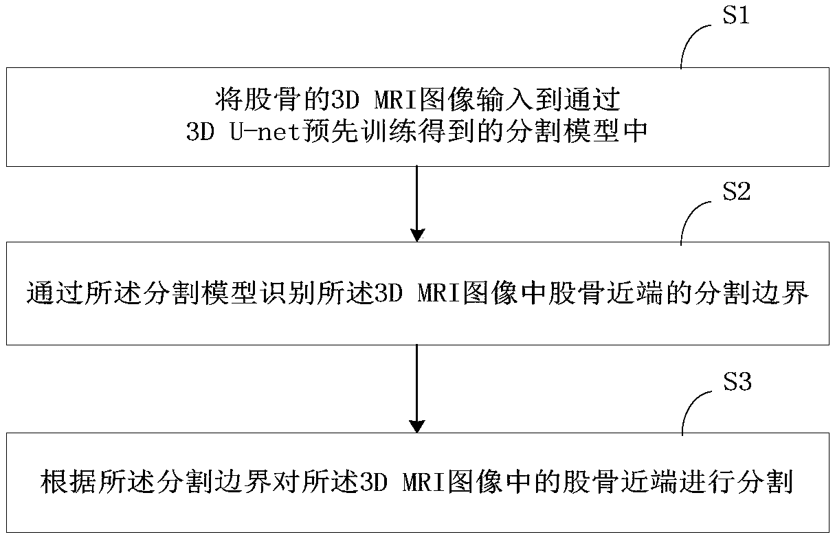
[0051] refer to figure 1 , the method for segmenting the proximal femur of an embodiment of the present application, comprising:

[0052] S1: Input the 3D MRI image of the femur into the segmentation model pre-trained by 3D U-net.

[0053]The 3D MRI (Magnetic Resonance Imaging, Image Magnetic Resonance Imaging) of the femur of the present embodiment is a "digital image" that is spatially coded by nuclear magnetic resonance signals. The magnetic resonance signals directly come from the object itself, and the magnetic resonance imaging can obtain any direction of the object...


PUM


Abstract

The invention discloses a proximal femur segmentation method and device, a computer device and a storage medium. The proximal femur segmentation method comprises the following steps: inputting a 3D MRI image of the femur into a segmentation model obtained by the pre-training of a 3D U-net; identifying the segmentation boundary of the proximal femur in the 3D MRI image through the segmentation model; segmenting the proximal femur in the 3D MRI image according to the segmentation boundary. According to the invention, the proximal femur is automatically separated from the 3D MRI image through thesegmentation model, and the proximal femur is separated from the original map, so that the diagnosis interference information can be reduced, and the diagnosis efficiency of a doctor can be greatly improved; a 3D MRI proximal femur segmentation technology based on the 3D U-net is provided, by means of the 3D U-net network with the depth monitoring and learning effects, the precise segmentation model is obtained through a small number of labeled samples, the accurate segmentation of the proximal femur of the 3D MRI proximal femur is achieved, and the technical problem that accurate segmentation is difficult due to the poor marked 3D MRI image data in the prior art is made up.

Description

technical field [0001] The present application relates to the field of computer technology, in particular to a method, device, computer equipment and storage medium for segmenting the proximal end of the femur. Background technique [0002] In recent years, due to the improvement of computer hardware level, deep neural network technology has been developed by leaps and bounds. Deep neural network technology has achieved great results in image, text or audio processing, but it has not been widely used in the medical field. Femoroacetabular impingement is one of the causes of hip pain in adults and is considered a key factor in cartilage damage and a precursor to osteoarthritis. MRI diagnosis is now used as a standard tool in the diagnosis of femoroacetabular impingement because it does not pose a radiological hazard. However, in the prior art, doctors generally conduct manual diagnosis by analyzing 2D MRI slice images based on medical experience, which has low diagnostic eff...

Claims


Application Information

Patent Timeline
Patent Type & Authority Applications(China)
IPC IPC(8): G06K9/34G06N3/04
CPCG06V10/267G06V2201/033G06N3/045
Inventor 王健宗吴天博刘新卉刘莉红马进肖京
Owner PING AN TECH (SHENZHEN) CO LTD
Why Eureka
Try Eureka