Quantitative description method of PET image nasopharyngeal carcinoma intra-tumor heterogeneity

A technology for nasopharyngeal carcinoma and heterogeneity, applied in the field of medical image analysis, can solve the problems that limit the adequacy and comprehensiveness, lack of specificity, and dependence of quantitative description of intratumoral heterogeneity, and achieve richness, adequacy and comprehensiveness Effect

Inactive Publication Date: 2018-11-13
SOUTHERN MEDICAL UNIVERSITY
View PDF4 Cites 3 Cited by
  • Summary
  • Abstract
  • Description
  • Claims
  • Application Information

AI Technical Summary

Problems solved by technology

18 F-FDG PET / CT has been widely used in the examination of nasopharyngeal carcinoma. However, because benign inflammatory lesions usually also show high glucose uptake, radiologists cannot observe the metabolic intensity and anatomical shape of lesions with naked eyes. Quantitative characterization of intratumoral heterogeneity image regions
Therefore, the identification of early nasopharyngeal carcinoma is easily interfered by inflammatory lesions, resulting in a specificity of less than 85%, and its diagnosis still relies on traumatic histopathological biopsy
However, the prior art methods for constructing texture matrices are relatively simple, and do not fully consider the influence of parameter settings such as symmetry, averaging strategy, distance, number of neighbors, and window width in the process of matrix construction, which greatly limits the texture matrix. Sufficiency and comprehensiveness of quantitative characterization of intraheterogeneity

Method used


Image

  • Quantitative description method of PET image nasopharyngeal carcinoma intra-tumor heterogeneity
  • Quantitative description method of PET image nasopharyngeal carcinoma intra-tumor heterogeneity
  • Quantitative description method of PET image nasopharyngeal carcinoma intra-tumor heterogeneity

Examples


Embodiment 1

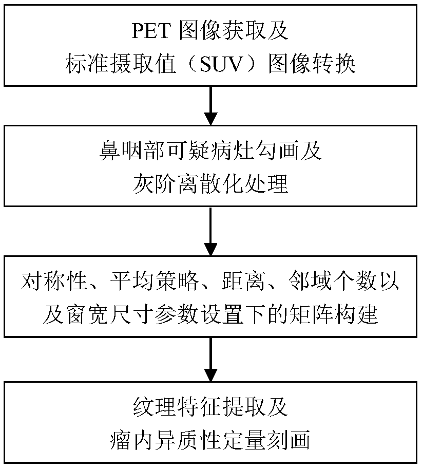
[0032] A quantitative characterization method for intratumoral heterogeneity of nasopharyngeal carcinoma in PET images, such as figure 1 shown, including:

[0033] The first step is to acquire the original image in DICOM format of the PET / CT scan of the target object, and perform preprocessing on the original image.

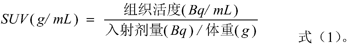
[0034] Due to the low resolution and small image size of PET images, usually 128*128, it is necessary to interpolate PET images in DICOM format to register and fuse them with CT images (512*512), and cubic splines can be used interpolation. In order to obtain the standard uptake value of each tissue, it is also necessary to convert the reconstructed attenuation-corrected activity map into a standard uptake value SUV image. The preprocessing of the original image in the present invention includes: interpolating the PET image, and combining with CT Image registration and fusion, and use the formula (1) to convert the PET activity map into a standard uptake value ...

Embodiment 2

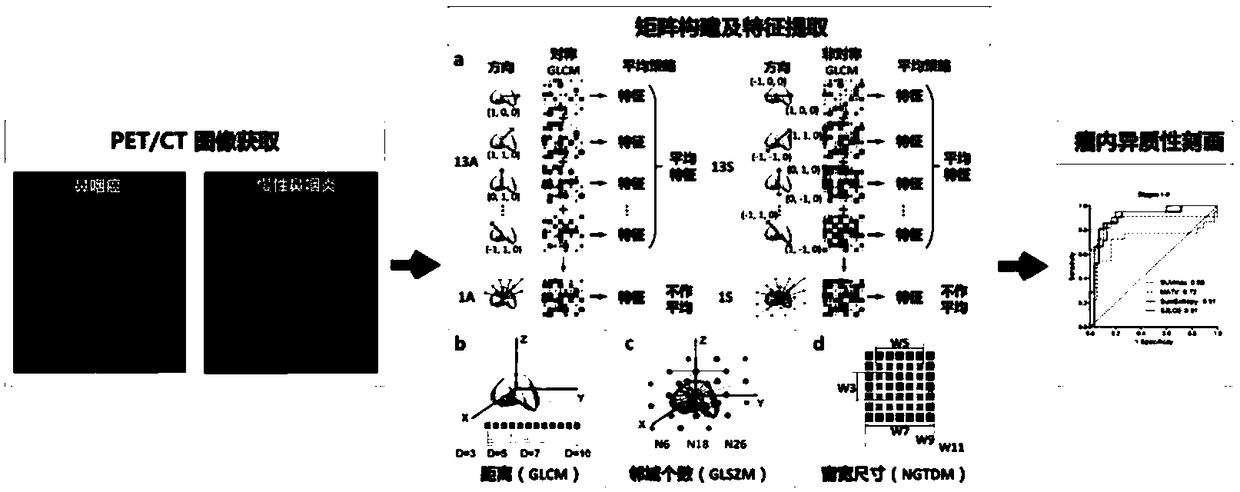
[0072] A quantitative characterization method for intratumoral heterogeneity of nasopharyngeal carcinoma in PET images, such as figure 2 shown, including the following steps.

[0073] First, the acquired DICOM format PET image was subjected to cubic spline interpolation, and it was registered and fused with the CT image to obtain an image size of 512*512 and a voxel size of 0.98*0.98*3. The following formula was used to convert the PET activity map into a standard uptake value SUV image:

[0074]

[0075] Then, 106 cases (69 cases of nasopharyngeal carcinoma and 37 cases of chronic nasopharyngitis) were segmented by using the threshold segmentation method with SUV>2.5 combined with the manual adjustment method.

[0076] The segmented lesions were discretized by SUV gray scale according to the following formula (SUV=0.1 for discrete interval):

[0077]

[0078] Among them, SUV(x) represents the standard uptake value of voxel x, B represents the discretization distance,...


PUM


Abstract

The invention belongs to the field of the medical image analysis, specifically a quantitative description method of PET image nasopharyngeal carcinoma intra-tumor heterogeneity. The method comprises the following steps: acquiring PET/CT scanning data of a patient, and converting the PET/CT scanning data into a standard SUV image; performing three-dimensional segmentation on the primary focus at the nasopharynx; performing discretization processing on the focus, and constructing four image omics matrixes for the processed focus, and comprehensively considering five parameter settings of symmetry, the average policy, the distance, the neighborhood number and window width size in the matrix construction process; extracting 415 texture features from the constructed matrix, and realizing the quantitative description of the intra-tumor metabolic heterogeneity by combining five traditional SUV features. The intra-tumor metabolic heterogeneity is described more comprehensively by sufficientlymining the information contained in the non-invasive PET image, thereby providing the quantitative evaluation index for precisely identifying the nasopharyngeal carcinoma by the doctor.

Description

technical field [0001] The invention relates to the technical field of medical image analysis, in particular to a method for quantitatively describing intratumoral heterogeneity of nasopharyngeal carcinoma in PET images. Background technique [0002] Nasopharyngeal carcinoma is an endemic disease that occurs mostly in southern China, with up to 30 cases per 100,000 people. 18 F-FDG PET / CT has been widely used in the examination of nasopharyngeal carcinoma. However, because benign inflammatory lesions usually also show high glucose uptake, radiologists cannot observe the metabolic intensity and anatomical shape of the lesion with naked eyes. Realize the quantitative description of intratumoral heterogeneity image area. Therefore, the identification of early nasopharyngeal carcinoma is easily interfered by inflammatory lesions, resulting in less than 85% specificity, and its diagnosis still depends on traumatic histopathological biopsy. Texture features have demonstrated the...

Claims


Application Information

Patent Timeline
Patent Type & Authority Applications(China)
IPC IPC(8): G06T7/136G06T7/41
CPCG06T2207/30096G06T7/136G06T7/41
Inventor 路利军吕闻冰马建华冯前进陈武凡
Owner SOUTHERN MEDICAL UNIVERSITY
Why Eureka
Try Eureka