Transcranial three-dimensional cerebrovascular compound imaging method and system

A composite imaging, cerebrovascular technology, applied in the IT and medical fields, can solve the problems of being unsuitable for multiple detections, limited clinical applications, and radiation, to achieve 3D scanning and long-term monitoring, and improve spatial form and position accuracy. and signal strength, the effect of convenient wearing

Inactive Publication Date: 2018-11-23
SHENZHEN DELICA MEDICAL EQUIP CO LTD
View PDF8 Cites 13 Cited by
  • Summary
  • Abstract
  • Description
  • Claims
  • Application Information

AI Technical Summary

Problems solved by technology

DSA is the gold standard for the detection of cerebrovascular lesions. CTA is more commonly used, but it is irradiated, invasive, and requires contrast agents. It is not suitable for multipl

Method used


Image

  • Transcranial three-dimensional cerebrovascular compound imaging method and system
  • Transcranial three-dimensional cerebrovascular compound imaging method and system
  • Transcranial three-dimensional cerebrovascular compound imaging method and system

Examples


Embodiment 1

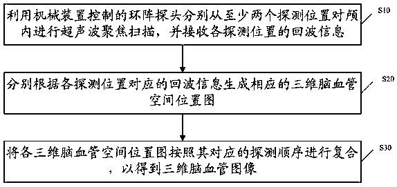
[0022] The application provides a transcranial three-dimensional cerebrovascular composite imaging method, such as figure 1 As shown, the method includes:

[0023] S10 , using a circular array probe controlled by a mechanical device to perform focused ultrasonic scanning of the intracranial from at least two detection positions, and receive echo information from each detection position.

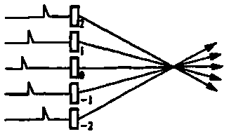
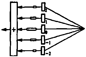
[0024] Specifically, the ring array probe controlled by the mechanical device may include a mechanical device and a ring array transducer, the mechanical device is used to drive the mechanical structure of the ring array probe to swing, and the ring array transducer is used to The focused ultrasonic signal is transmitted internally, and the probe that receives the echo signal of the ultrasonic signal is focused. In this embodiment, the ring array transducer includes several element chips arranged in concentric circles, and the area of ​​each element chip is equal. For example, the ring arra...

Embodiment 2

[0067] This embodiment provides a transcranial three-dimensional cerebrovascular composite imaging method. This implementation is the same as the imaging process in Embodiment 1, the difference lies in the acquisition process of the echo data of each scanning position by the ring array probe, so here The acquisition process of the echo data at each scanning position by the ring array probe is described in detail. The ring array probe data acquisition process may specifically include:

[0068] A. Adjust the probe angle and scanning depth according to the preset swing trajectory, and send multi-beam focused ultrasonic signals to the intracranial at the scanning position, respectively receive the echo signals of the multi-beam ultrasonic signals and record the echo signals of the scanning position and each scanning depth ;

[0069] B. Send multiple beams of ultrasonic signals at the scanning position and receive echo signals of multiple ultrasonic signals, and retain the stronge...


PUM


Abstract

The invention discloses a transcranial three-dimensional cerebrovascular compound imaging method and system. The method comprises the steps that firstly, an annular array probe capable of performing automatic scanning is utilized to send ultrasonic signals with different time delays to intracranial different scanning positions, so that the ultrasonic signals are focused on preset depths of the beam axial direction to detect the intracranial cerebral vessels through the focused ultrasonic signals and perform digital delay focusing on received echo signals to enhance the echo signals to improvethe spatial form, and positional accuracy of acquired cerebral vessels, thereby improving the definition and detection rate of three-dimensional cerebrovascular images. In addition, the three-dimensional cerebrovascular images are acquired from different detection positions, according to the three-dimensional cerebrovascular images, intracranial cerebral vessels are compounded from multiple angles, thereby further improving the definition of the three-dimensional cerebrovascular images. The annular array probe has the characteristics of small size and low frequency, so that the annular array probe can be worn on the head conveniently, and three-dimensional scanning and long-time monitoring of the intracranial cerebral vessels can be achieved.

Description

technical field [0001] The invention relates to the fields of IT and medical technology, in particular to a transcranial three-dimensional cerebrovascular composite imaging method and system. Background technique [0002] Stroke is the number one cause of death in China. There is an urgent and huge need to use safe and effective equipment to diagnose patients with cerebrovascular diseases and to screen high-risk groups for stroke. The existing color Doppler ultrasound diagnostic equipment (TCCD) has clear and high-resolution diagnostic images of intracranial blood vessels, but due to the huge attenuation of the skull to ultrasound, the detection rate is low, the operation is difficult, and it is dependent on personnel or equipment powerful. Ultrasound transcranial Doppler (TCD) has a high detection rate, but there is no two-dimensional image, and the operation is blind typing, which is highly dependent on personnel. DSA is the gold standard for the detection of cerebrovas...

Claims


Application Information

Patent Timeline
IPC IPC(8): A61B8/06
CPCA61B8/06A61B8/5207A61B8/5215
Inventor 杨弋邢英琦王筱毅周果欧阳俊华梁志成
Owner SHENZHEN DELICA MEDICAL EQUIP CO LTD
Why Eureka
Try Eureka