

MRI t2 image imaging method
A nuclear magnetic resonance and image imaging technology, applied in magnetic resonance measurement, measurement of magnetic variables, measurement of magnetic variables, etc., can solve problems such as inability to apply
- Summary
- Abstract
- Description
- Claims
- Application Information
AI Technical Summary
Problems solved by technology
Method used
Image

Examples
Embodiment Construction
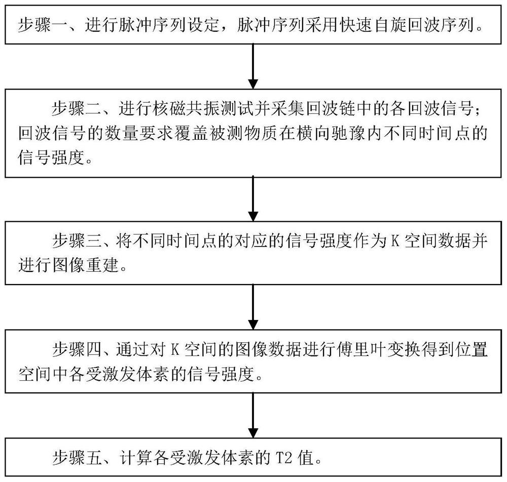
[0039] Such as figure 1 As shown, it is a flow chart of the method of the embodiment of the present invention, and the nuclear magnetic resonance T2 image imaging method of the embodiment of the present invention includes the following steps:
[0040] Step 1. Perform pulse sequence setting, and the pulse sequence adopts a fast spin echo sequence.
[0041] The fast spin echo sequence is a single shot fast spin echo.
[0042] The fast spin echo sequence includes: one excitation radio frequency pulse for excitation and multiple subsequent refocusing radio frequency pulses for flipping and refocusing, each refocusing radio frequency pulse corresponds to a spin echo, each of the Spin echoes form one of the echo signals that are detected.
[0043] The excitation radio frequency pulse is a radio frequency pulse with an angle of 70°-120° to invert the magnetization vector.
[0044] Each of the refocusing radio frequency pulses is a radio frequency pulse that reverses the magnetizat...
PUM
 Abstract
Description
Claims
Application Information