Left-atrial-appendage CT image segmentation method

A CT image and image technology, applied in the field of medical image processing, can solve the problems of poor effect and the inability to directly obtain the boundary of the left atrial appendage CT image.

Inactive Publication Date: 2018-11-23
XIAMEN UNIV
View PDF4 Cites 11 Cited by
  • Summary
  • Abstract
  • Description
  • Claims
  • Application Information

AI Technical Summary

Problems solved by technology

CT images of the left atrial appendage have the following characteristics: there is no border between the left atrial appendage and the left atrium, and the parenchyma of the left atrial appendage has no texture features. Therefore, the border of the CT image of the left atrial appendage cannot be directly obtained, and the traditional image segmentation algorithm is not effective.

Method used


Image

  • Left-atrial-appendage CT image segmentation method
  • Left-atrial-appendage CT image segmentation method
  • Left-atrial-appendage CT image segmentation method

Examples


Embodiment Construction

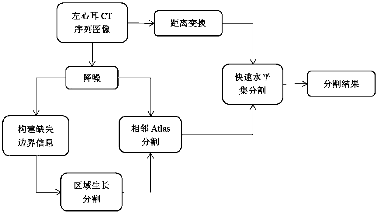
[0082] The present invention will be described in further detail below in conjunction with accompanying drawing and embodiment, embodiment according to figure 1 The flowchart shown is explained in detail.

[0083] The present invention comprises the following steps:

[0084] 1) Input the left atrial appendage CT sequence image A;

[0085] 2) Carry out denoising processing to sequence image A in step 1), obtain sequence image B;

[0086] 3) Select a single CT image B in the sequence image B in step 2) i , and the remaining sequence images are denoted as B';

[0087] 4) According to the image B obtained in step 3) i For the location feature information of the middle left atrial appendage, manually select two feature points to construct the missing boundary information of the left atrial appendage, and obtain image C;

[0088] 5) Use the region growing algorithm on the image C obtained in step 4) to obtain the image L i ;

[0089] 6) Image L obtained in step 5) i To initi...


PUM


Abstract

The invention discloses a left-atrial-appendage CT image segmentation method, and relates to medical image processing. Firstly, left-atrial-appendage CT images are denoised, prior knowledge is utilized to construct missing boundary information of a left atrial appendage, and a region growing algorithm is used to obtain a single initial segmentation image to use the same as a single Atlas label graph; then information relationships of the single label graph and an adjacent left-atrial-appendage CT sequence are utilized for improved Atlas segmentation to obtain an initial segmentation sequence;contour information in the initial segmentation sequence is used as initial contours of a level set algorithm, and approximate Euclidean distance information of left-atrial-appendage CT sequence graphs is obtained to be used as feature input; and finally, a precise segmentation result sequence is obtained through a fast level set algorithm to achieve the purpose of left-atrial-appendage CT image segmentation.

Description

technical field [0001] The invention relates to medical image processing, in particular to a left atrial appendage CT image segmentation method. Background technique [0002] The left atrial appendage is a part of the left atrium of the human body, and its function is to assist in pumping blood. The multi-branched structure of the left atrial appendage is easy to hinder blood return. When the left atrial appendage cannot produce normal contraction, it will increase the pressure of the atrium and cause atrial fibrillation. The structure of the left atrial appendage has an important impact on cardiovascular and cerebrovascular diseases. When performing occlusion surgery on the left atrial appendage, how to choose a suitable occlusion device according to the shape of the left atrial appendage is the basis for the success of the operation. Therefore, left atrial appendage CT image segmentation is one of the foundations for left atrial appendage image analysis and processing, wh...

Claims


Application Information

Patent Timeline
Patent Type & Authority Applications(China)
IPC IPC(8): G06T7/00G06T7/11G06T7/136G06T7/187
CPCG06T7/0012G06T7/11G06T7/136G06T7/187G06T2207/10081G06T2207/20156G06T2207/30048
Inventor 黄晓阳张耀丹苏茂龙
Owner XIAMEN UNIV
Why Eureka
Try Eureka