
An Automatic Liver Segmentation Method for Abdominal CT Sequence Images Based on Level Set and Shape Descriptor

A shape description and sequence image technology, applied in the field of medical image analysis and processing, can solve the problems of unsatisfactory low-contrast CT image segmentation results, noise sensitivity, and long training time, so as to improve liver segmentation accuracy, optimize liver edges, reduce effect of influence

Active Publication Date: 2022-03-29
湖南提奥医疗科技有限公司
View PDF11 Cites 0 Cited by
  • Summary
  • Abstract
  • Description
  • Claims
  • Application Information

AI Technical Summary

Problems solved by technology

Traditional methods are sensitive to noise, and the segmentation results of low-contrast CT images are not ideal
Shape model-based methods fail to accurately segment abnormally shaped livers
Liver segmentation based on deep learning requires a large amount of data as support during network training, which takes a long time to train and requires high hardware requirements

Method used


Image

  • An Automatic Liver Segmentation Method for Abdominal CT Sequence Images Based on Level Set and Shape Descriptor
  • An Automatic Liver Segmentation Method for Abdominal CT Sequence Images Based on Level Set and Shape Descriptor
  • An Automatic Liver Segmentation Method for Abdominal CT Sequence Images Based on Level Set and Shape Descriptor

Examples


Embodiment 1

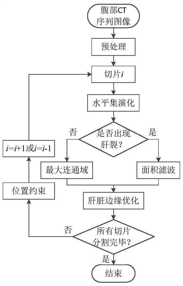
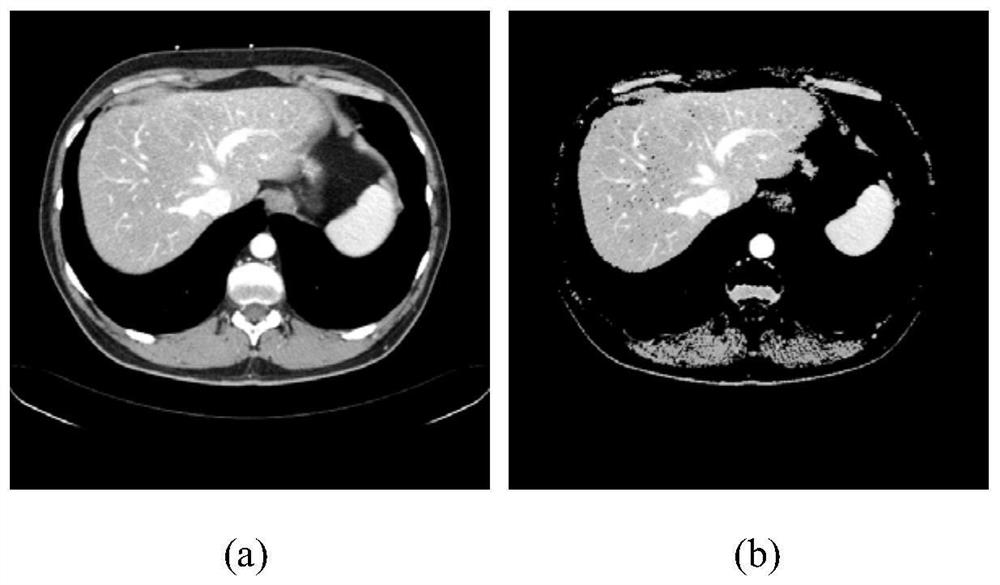
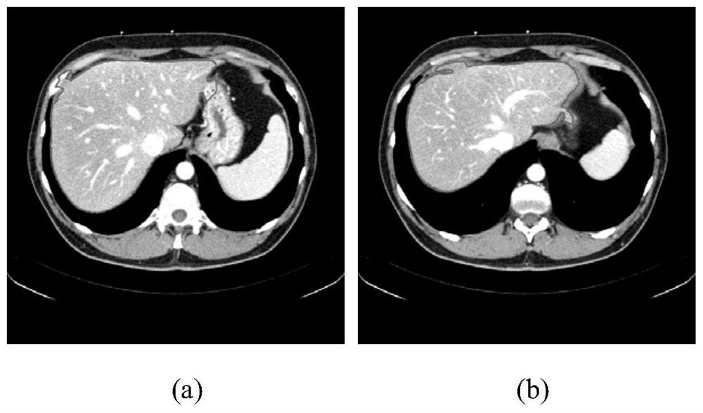
[0032] figure 1 Shown is a flow chart of a method for automatic liver segmentation of abdominal CT sequence images based on level sets and shape descriptors according to an embodiment of the present invention. Firstly, the input abdominal CT sequence image is preprocessed; then, the initial liver slice is selected, and the liver is initially segmented using the level set method incorporating the gray offset field; then, the edge of the initial liver slice is optimized; finally, the initial liver slice is Slices are used as the starting point, and the liver segmentation results of adjacent slices are incorporated into the level set energy function as position constraints, and all slices are iteratively segmented up and down.

[0033] Combine below figure 1 , using a preferred embodiment to describe in detail the method for automatic liver segmentation of abdominal CT sequence images based on level sets and shape descriptors of the present invention.

[0034] 1. Pretreatment. ...

Embodiment 2

[0048] The Sliver07 and XHCSU14 databases were tested using the method in Example 1. The Sliver07 database contains 20 abdominal CT sequences from different patients, the slice image size is 512×512, the plane pixel spacing is distributed in the range of 0.5762mm to 0.8125mm, and the slice thickness is distributed in the range of 0.7mm to 3.0mm; XHCSU14 database Provided by Xiangya Hospital of Central South University, the database contains 20 abdominal CT sequences from different patients. The slice image size is 512×512, the plane pixel pitch ranges from 0.5313mm to 0.7402mm, and the slice thickness is 1.0mm and 1.5mm . Volume Overlap Error (Volumetric Overlap Error, VOE), Relative Volume Difference (Relative Volume Difference, RVD), Average Symmetric Surface Distance (Average Symmetric Surface Distance, ASD), Root Mean Square Symmetric Surface Distance (Root Mean Square Symmetric Surface Distance, RMSD) and Maximum Symmetric Surface Distance (MaximumSymmetric Surface Dista...


PUM


Abstract

The invention discloses a method for automatic liver segmentation of abdominal CT sequence images based on level set and shape descriptor, which includes: preprocessing the input image to remove irrelevant organs and tissues; The correlation between adjacent slices in the sequence was used to construct a level set energy function. Starting from the initial slice, an iterative strategy was used to complete the automatic liver segmentation of abdominal CT sequence images; local and global shape descriptors were constructed to remove over-segmented regions and optimize the liver edge. The method of the present invention can effectively segment the liver area in the abdominal CT sequence image which is seriously polluted by noise and has gray scale heterogeneity, can effectively avoid wrong segmentation of adjacent tissues around the liver, remove over-segmented areas caused by gray scale overlap, and improve Liver segmentation accuracy.

Description

technical field [0001] The invention relates to the field of medical image analysis and processing, in particular to an automatic liver segmentation method for abdominal CT sequence images based on level sets and shape descriptors. Background technique [0002] my country is a country with a large number of liver diseases, and more than half of the new cases of liver diseases and deaths due to liver diseases in the world occur in China. Currently, the main treatment methods for liver diseases include liver resection, living donor liver transplantation, and stereotactic radiotherapy. Computer-aided diagnosis and surgical planning are an important part of the treatment of liver diseases. Accurate liver image segmentation is the basis of computer-aided diagnosis and surgical planning, and can provide technical support for liver lesion analysis, surgical navigation, and radiotherapy planning. Due to the large number of slices in abdominal CT sequence images, it is time-consumi...

Claims


Application Information

Patent Timeline
Patent Type & Authority Patents(China)
IPC IPC(8): G06T7/11
CPCG06T7/11G06T2207/30056G06T2207/10016G06T2207/10081
Inventor 赵于前李阳廖苗廖胜辉杨振
Owner 湖南提奥医疗科技有限公司
Learn More