Complex capable of inhibiting genetic function in exosome, and cancer proliferation and/or metastasis suppressor

An inhibitor, exosome technology, applied in the field of genetically functional conjugates

Inactive Publication Date: 2018-12-21
KYOTO UNIV +1
View PDF3 Cites 2 Cited by
  • Summary
  • Abstract
  • Description
  • Claims
  • Application Information

AI Technical Summary

Problems solved by technology

However, since miRNAs in blood are encapsulated in exosomes, it is difficult to directly target miRNAs even by administering antisense nucleic acids into blood

Method used


Image

  • Complex capable of inhibiting genetic function in exosome, and cancer proliferation and/or metastasis suppressor
  • Complex capable of inhibiting genetic function in exosome, and cancer proliferation and/or metastasis suppressor
  • Complex capable of inhibiting genetic function in exosome, and cancer proliferation and/or metastasis suppressor

Examples


Embodiment 1

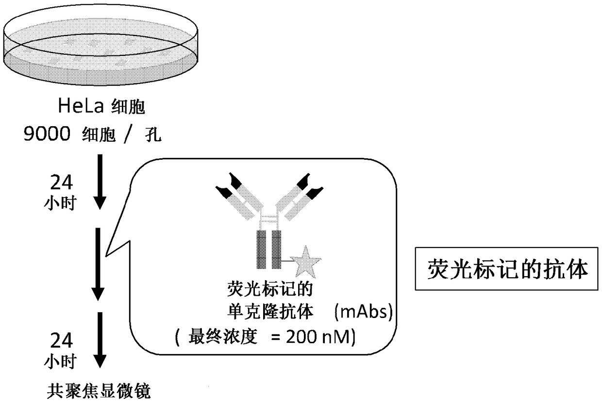
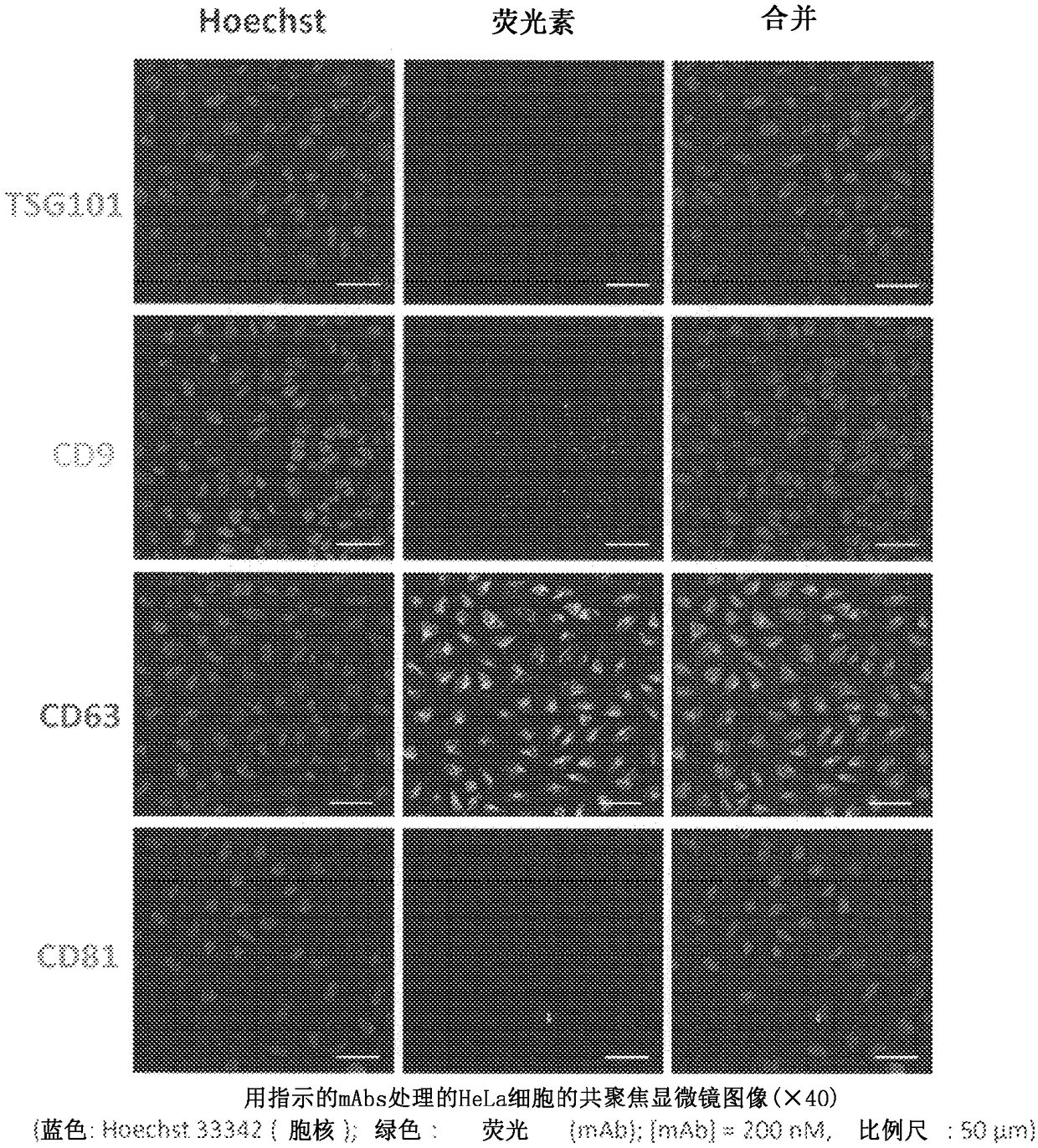
[0066] Example 1: Capturing fluorescently labeled antibodies into cells ( figure 2 )

[0067] It was verified whether antibodies recognizing exosome surface antigens were incorporated into cells together with exosomes.

[0068] Hela cells (cervical cancer cells) were inoculated onto porous glass-bottomed dishes (Matsunami GlassInd., Ltd) at an amount of 9000 / well in 5% CO 2 Incubator at 37°C for 24 hours. Add fluorescently labeled antibodies (anti-CD63 antibody, anti-CD9 antibody, anti-CD81 antibody, and anti-TSG101 antibody) to each well, and then use 5% CO 2 The incubator was incubated at 37°C for 24 hours. Remove the supernatant and wash the cells with 1X PBS. Add 100 μL of 4% paraformaldehyde followed by incubation for 5 min at room temperature to fix the cells. Wash cells twice with 1X PBS. Viable cells were stained by adding 200 μL of Hoechst 33342 in 1×PBS (final concentration=5 μM), followed by incubation at room temperature for 10 minutes. Cells were washed tw...

Embodiment 2 and comparative example 1

[0071] Example 2 and Comparative Example 1: Capturing antibody / nucleic acid conjugates into cells ( Figure 4 )

[0072] It was verified whether antibodies recognizing exosome surface antigens were incorporated into cells together with exosomes.

[0073] Hela cells (cervical cancer cells) were inoculated onto porous glass-bottomed dishes (Matsunami GlassInd., Ltd) at an amount of 9000 / well in 5% CO 2 Incubator at 37°C for 24 hours. An anti-CD63 antibody-9r / nucleic acid conjugate (anti-CD63 IgG-9r+anti-miR(Cy5)) was added to each well. The anti-miR(Cy5) used here was 5'-Cy5-aguca auagggugug ugaga gacuu acug-3' (FASMAC, SEQ ID NO: 1).

[0074] As Comparative Example 1, anti-CD63 IgG and anti-miR (Cy5) were added instead of the anti-CD63 IgG-9r / nucleic acid conjugate.

[0075] Phalloidin was used to stain the cytoskeleton. Phalloidin is an oligopeptide that specifically binds to the polymeric actin (F-actin) that makes up the cytoskeleton.

[0076] Use 5% CO 2 Incubator at 3...

Embodiment 3

[0077] Example 3: MicroRNA function inhibitory effect of anti-CD63 antibody / anti-miR nucleic acid conjugate ( Figure 6 )

[0078] The inhibitory effect of microRNA function upon incorporation of anti-CD63 antibody / anti-miR nucleic acid conjugates into cells was assessed.

[0079] Hela cells (cervical cancer cells) were seeded on 96-well plates at an amount of 4500 / well, and 5% CO was used to 2 Incubator at 37°C for 24 hours. MicroRNA (miR-Luc) targeting luciferase mRNA was introduced into each well (LipofectamineRNAiMAX). Use 5% CO 2 The incubator was incubated at 37°C for 18 hours, and luciferase-expressing plasmids (pGL4.13 and pGL4.73) were introduced (Lipofectamine 2000). Use 5% CO 2 The incubator was incubated at 37°C for 6 hours, and anti-CD63 antibody / anti-miR nucleic acid conjugate (anti-CD63 IgG-9r+anti-miR-Luc), anti-miR-Luc only (300nM) or anti-CD63 antibody only (600nM) was added, and use 5% CO 2 The incubator was incubated at 37°C for 24 hr; firefly lucife...


PUM

PropertyMeasurementUnit
diameteraaaaaaaaaa

Abstract

The present invention provides a complex comprising an antibody that targets an exosome surface antigen or an antibody fragment of the antibody and a suppressor of a gene or an expression product of the gene, wherein the antibody or the antibody fragment and the suppressor of the gene or the expression product of the gene are covalently bound to each other directly or through a linker or are non-covalently bound to each other.

Description

technical field [0001] The present invention relates to conjugates capable of inhibiting the genetic function of exosomes, and inhibitors of cancer proliferation and / or metastasis. Background technique [0002] Recent research has discovered a mechanism in which various cells, including T cells, platelets, epithelial cells, immune cells or cancer cells, release vesicles called exosomes with a diameter of 40 to 100 nm to transmit information to distant cells. cells. [0003] Specifically, a novel mechanism in which cancer cell proliferation and metastatic ability is dominated by the expression products of genes (such as miRNAs) contained in exosomes secreted in blood has been proposed, and this mechanism has caused great focus on. [0004] In order to inhibit the function of miRNA in exosomes, a method using a modified nucleic acid (antisense nucleic acid) having a sequence complementary to miRNA is generally used (Non-Patent Documents 1 to 3). However, since miRNAs in blo...

Claims


Application Information

Patent Timeline
Patent Type & Authority Applications(China)
IPC IPC(8): A61K39/395A61K31/7105A61K48/00A61P35/00A61P35/04
CPCA61K47/6849A61K47/6883A61K31/7105A61K31/713A61P35/00A61P35/04C07K16/2803C07K16/2896A61K2039/505C07K2317/77A61K47/6455C12N15/113C12N15/87C12N15/111C12N2310/113C12N2320/31A61K2300/00A61K47/549
Inventor 山吉麻子村上章芦原英司小堀哲生
Owner KYOTO UNIV
Why Eureka
Try Eureka