
A method for segmentation of pulmonary nodules in lung CT images

A technology for CT images and pulmonary nodules, applied in image analysis, image enhancement, image data processing, etc., can solve problems such as misdiagnosis or missed diagnosis, difficult to detect diseases, and radiologists' energy, etc. The effect of improving accuracy and simple algorithm

Inactive Publication Date: 2019-01-15
ZHENGZHOU UNIV
View PDF5 Cites 14 Cited by
  • Summary
  • Abstract
  • Description
  • Claims
  • Application Information

AI Technical Summary

Problems solved by technology

[0002] Lung cancer is the cause of death among cancers with a high incidence, because its early asymptomatic display makes the disease difficult to detect. When the disease is discovered, the cancer has already metastasized, which makes lung cancer difficult to treat. Lung cancer often has symptoms of lung nodules in the early stage. Chest CT scans are mostly used for nodule detection, and the scan results are characterized by clear and accurate images. However, a patient’s test results will have hundreds of pictures, and it takes a lot of effort for radiologists to find them. At the same time, repeated work for a long time may also cause Misdiagnosis or missed diagnosis

Method used


Image

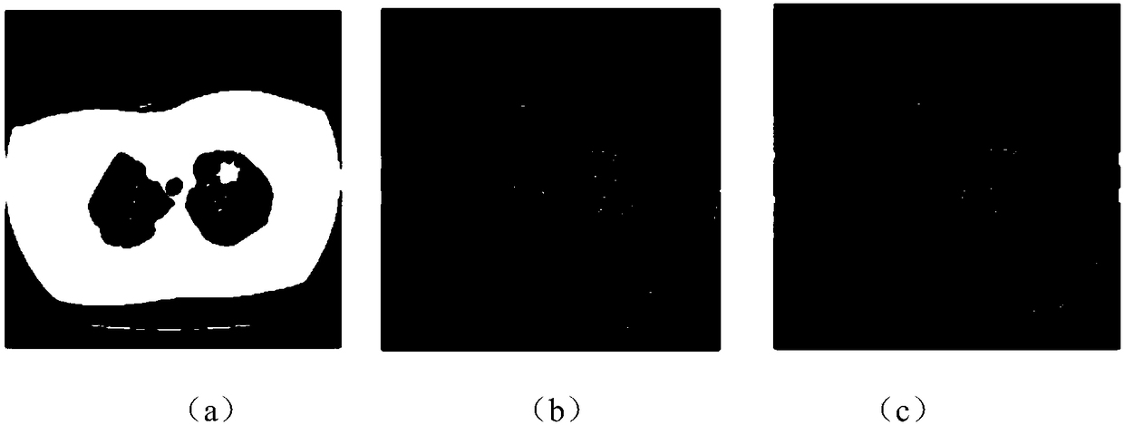
  • A method for segmentation of pulmonary nodules in lung CT images
  • A method for segmentation of pulmonary nodules in lung CT images
  • A method for segmentation of pulmonary nodules in lung CT images

Examples


Embodiment Construction

[0041] The following will clearly and completely describe the technical solutions in the embodiments of the present invention with reference to the accompanying drawings in the embodiments of the present invention. Obviously, the described embodiments are only some, not all, embodiments of the present invention. Based on the embodiments of the present invention, all other embodiments obtained by persons of ordinary skill in the art without making creative efforts belong to the protection scope of the present invention.

[0042] The data used in the present invention is LIDC-IDRI, which is a data set composed of chest medical image files and corresponding diagnosis results, and the data set is initiated and collected by the National Cancer Institute, an American scientific research institute.

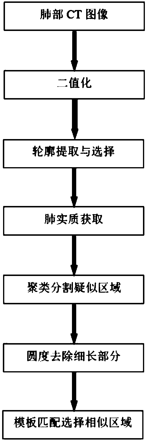
[0043] like figure 1 Shown, a lung nodule segmentation method in a lung CT image, comprising the following steps:

[0044] (1) Extract the lung parenchyma contour line including lung no...


PUM


Abstract

The invention provides a segmentation method of pulmonary nodules in lung CT images, comprising the following steps: (1) extracting pulmonary parenchyma contour lines including pulmonary nodules fromlung images to obtain pulmonary parenchyma images; (2) adopting clustering algorithm to segment the suspected pulmonary nodule region from the pulmonary parenchyma image in the step (1); (3) removingthe region of suspected pulmonary nodules by image roundness method, and eliminating the interference of blood vessel or trachea; (4) according to the size of the suspected pulmonary nodules in step (3) Gaussian template is constructed, and the region with high correlation coefficient is selected to extract the pulmonary nodules. The invention considers the shape characteristic of the solitary pulmonary nodule and the distribution characteristic of the image gray value formed by the change of the CT value of the lung, and reduces the error of the segmentation result of the pulmonary nodule.

Description

technical field [0001] The invention relates to the technical field of image segmentation, in particular to a method for segmenting pulmonary nodules in lung CT images. Background technique [0002] Lung cancer is the cause of death among cancers with a high incidence, because its early asymptomatic display makes the disease difficult to detect. When the disease is discovered, the cancer has already metastasized, which makes lung cancer difficult to treat. Lung cancer often has symptoms of lung nodules in the early stage. Chest CT scans are mostly used for nodule detection, and the scan results are characterized by clear and accurate images. However, a patient’s test results will have hundreds of pictures, and it takes a lot of effort for radiologists to find them. At the same time, repeated work for a long time may also cause misdiagnosis or missed diagnosis. [0003] Computer-aided diagnosis technology is a technology that assists doctors in diagnosing diseases by summari...

Claims


Application Information

Patent Timeline
Patent Type & Authority Applications(China)
IPC IPC(8): G06K9/46G06K9/62G06T7/11
CPCG06T7/11G06T2207/30064G06V10/44G06F18/23213
Inventor 董敏李舒意孙燚段鋆心穆晓敏
Owner ZHENGZHOU UNIV
Learn More