Power spectrum based ultrasonic scattering particle diameter imaging method

An imaging method and scatterer technology, applied in the field of signal processing, can solve problems such as missing frequency and limited diagnostic information of ultrasonic imaging, and achieve the effect of high precision and low computational complexity

Active Publication Date: 2019-01-22
SHENZHEN CERTAINN TECH CO LTD
View PDF6 Cites 2 Cited by
  • Summary
  • Abstract
  • Description
  • Claims
  • Application Information

AI Technical Summary

Problems solved by technology

The commonly used clinical "B-ultrasound" (B-mode ultrasound imaging) uses the amplitude information of the backscattered signal (rad...

Method used


Image

  • Power spectrum based ultrasonic scattering particle diameter imaging method
  • Power spectrum based ultrasonic scattering particle diameter imaging method
  • Power spectrum based ultrasonic scattering particle diameter imaging method

Examples


Embodiment Construction

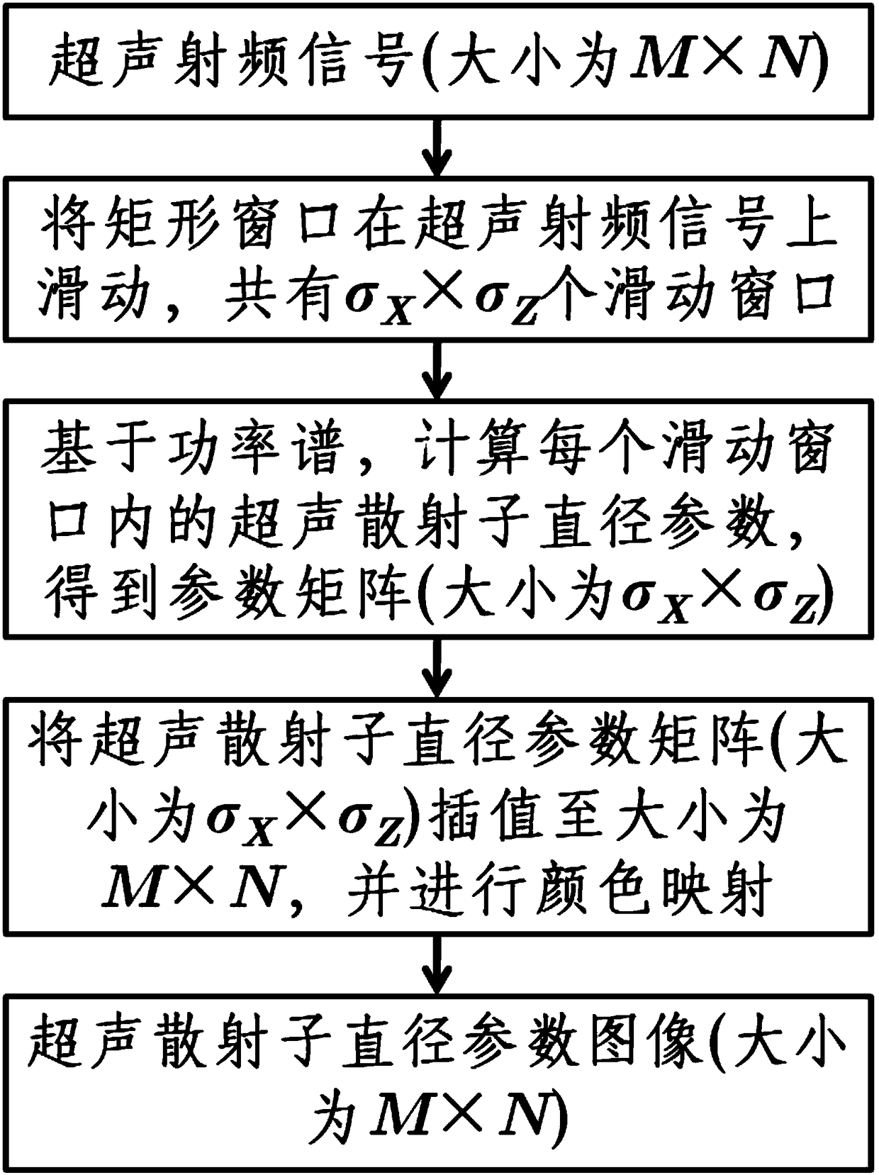
[0022] The ultrasonic scattering sub-diameter imaging method based on the power spectrum of the present invention is based on the ultrasonic backscattering signal (radio frequency signal) of the tissue to be measured, calculates the power spectrum, then calculates the ultrasonic scattering sub-diameter parameter and calculates the ultrasonic scattering sub-diameter parameter image method.

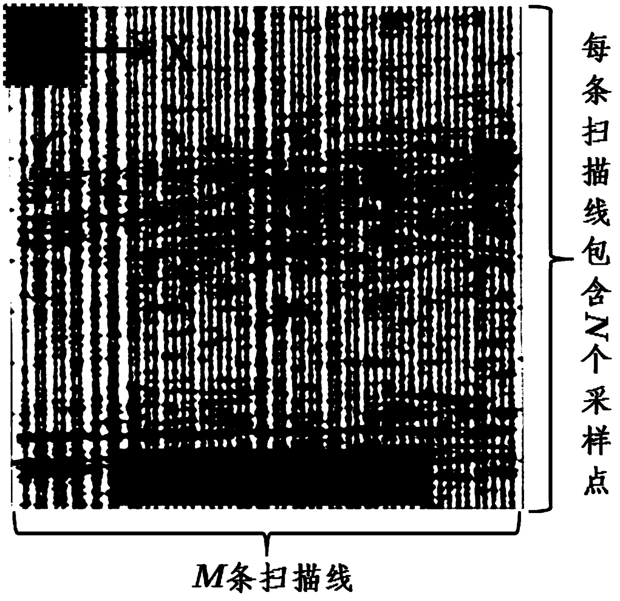
[0023] Without loss of generality, assuming that the ultrasonic radio frequency signal is composed of M scanning lines, and each scanning line contains N sampling points, the ultrasonic radio frequency signal is a two-dimensional matrix with a size of M×N. figure 1 Be the flowchart of the inventive method, mainly comprise the following steps:

[0024] (1) Slide the rectangular window on the ultrasonic RF signal, such as figure 2 As shown, the size of this sliding window is M w ×N w , which means M w scanning lines×N w sampling points. Let the sliding window be in the X and Z directio...


PUM


Abstract

The invention discloses a power spectrum based ultrasonic scattering particle diameter imaging method. Based on ultrasonic radio frequency signals, the method calculates a power spectrum, calculates an ultrasonic scattering particle diameter parameters and calculates an ultrasonic scattering particle diameter parameter image. The method is as below: sliding a sliding window over an ultrasonic RF signal; calculating ultrasonic scattering particle parameters in each sliding window based on the power spectrum to obtain a matrix of ultrasonic scattering particle parameter values; interpolating thematrix of the ultrasonic scattering sub-parameter values into the size of the ultrasonic RF signal; and performing color mapping to obtain an ultrasound scattering particle diameter parameter image.The ultrasonic scattering particle diameter imaging method of the present invention can be used for ultrasonic tissue characterization of biological tissues such as mammary gland, liver and the like.

Description

technical field [0001] The invention belongs to the technical field of signal processing, and in particular relates to a medical ultrasonic signal processing method, more specifically, a method for calculating ultrasonic scattering sub-parameters and imaging using the power spectrum of ultrasonic backscattering signals (radio frequency signals). Background technique [0002] Ultrasound imaging is widely used in clinical diagnosis due to its real-time performance, low cost, and no ionizing radiation. The ultrasonic probe (transducer) emits ultrasonic waves into the tissue, and after a series of interactions between the ultrasonic waves and the tissue such as scattering, reflection, and diffraction, the ultrasonic probe receives the backscattered echoes of the tissue. The commonly used clinical "B-ultrasound" (B-mode ultrasound imaging) uses the amplitude information of the backscattered signal (radio frequency signal) for imaging, but loses information such as frequency, resu...

Claims


Application Information

Patent Timeline
IPC IPC(8): A61B8/00
CPCA61B8/52
Inventor 周著黄欧阳亚丽吴水才高宏建丁琪瑛
Owner SHENZHEN CERTAINN TECH CO LTD
Why Eureka
Try Eureka