Device and method for removing free drug in anti-drug antibody detecting sample, preparation method and application thereof

A technology for antibody detection and drug application in the fields of biomedicine and sample pretreatment

Active Publication Date: 2019-02-19
SHANGHAI INST OF MATERIA MEDICA CHINESE ACAD OF SCI
View PDF10 Cites 2 Cited by
  • Summary
  • Abstract
  • Description
  • Claims
  • Application Information

AI Technical Summary

Problems solved by technology

2) The re-acidification step is to achieve the purpose of eluting the anti-drug antibodies bound to the micropore plate, but since the drug coated on the micropore plate is achieved by passive adsorption of non-covalent bonds, the acid During elution, not only the anti-drug antibody is eluted from the coated drug molecule, but also the complex of drug and anti-drug antibody is eluted, and the latter cannot be detected

Method used


Image

  • Device and method for removing free drug in anti-drug antibody detecting sample, preparation method and application thereof
  • Device and method for removing free drug in anti-drug antibody detecting sample, preparation method and application thereof
  • Device and method for removing free drug in anti-drug antibody detecting sample, preparation method and application thereof

Examples


preparation example Construction

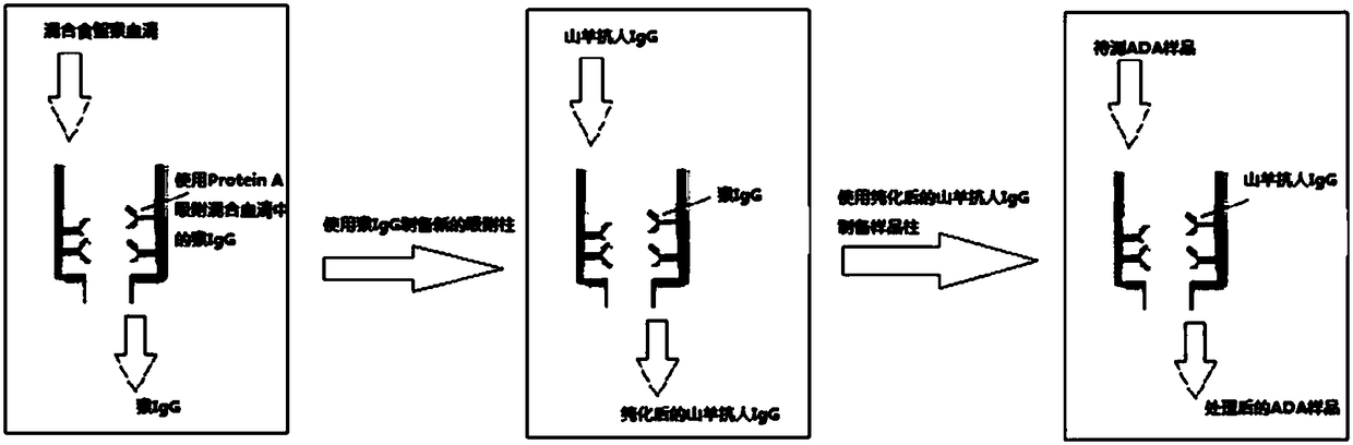
[0046] Preparation Example The preparation of adsorption column

[0047] First fill the column with Protein A+G (coupled with agarose), purify the monkey IgG in the monkey serum, elute the purified monkey IgG and collect it. The purified monkey IgG was reacted with NHS-activated agarose resin at room temperature for 1 h. After the reaction, the purification column was washed with PBS buffer, and the purification column was blocked with 1M Tris buffer (pH 7.4), and reacted at room temperature for 15-20 min. Then, the purification column was washed with PBS buffer to prepare monkey IgG-coupled agarose resin. Goat anti-human IgG polyclonal antibody (Beijing Biolab Technology Co., Ltd.; specification / model: F020227) was purified by monkey IgG-coupled agarose resin, and the purified goat anti-human IgG polyclonal antibody The anti-human IgG-coupled agarose resin was prepared by using the same method as the preparation method of the above-mentioned monkey IgG-coupled agarose resin,...

experiment Embodiment 1

[0110] Experimental Example 1 Evaluation of the Antibody Coupling Ability of Adsorption Cartridges

[0111] Through the above general ELISA method, it is found that the adsorption column (goat anti-human IgG) obtained in the preparation example can be combined with no less than 450 μg of human IgG under selected coupling conditions. The results are shown in Figure 4 .

[0112] After coupling the column with 400 μg of goat anti-human IgG, it was used to adsorb different amounts of human IgG (simulated drug) at room temperature for 1 hour. It was found that when the human IgG was 500 μg, the adsorption effect of the column reached 90% (see Figure 5 indicated by the arrow in).

[0113] Experimental example 2 Adsorption of the purified anti-PD-L1 humanized monoclonal antibody drug by the adsorption column obtained in the preparation example

[0114] After coupling the column with 400 μg of goat anti-human IgG, it was used to adsorb the purified anti-PD-L1 humanized monoclonal ...


PUM


Abstract

The invention provides a device and method for removing free drug in an anti-drug antibody detecting sample, a preparation method and application thereof. Specifically, a substance that specifically binds to a drug is coupled to a solid phase carrier, and does not react with other components in the sample other than the free drug in the device. The interference of the antibody drug on the anti-drug antibody detection can be effectively reduced, and the sensitivity of anti-drug antibody detection in the sample is improved by using the device of the invention to pretreat the anti-drug antibody detecting sample and detecting the anti-drug antibody in the sample in combination with the anti-drug antibody detecting method (such as a bridging method).

Description

technical field [0001] The invention belongs to the field of sample pretreatment in the field of biomedicine, in particular, it relates to a device and method for removing free drugs in a sample, a preparation method and application of the device, in particular, the invention relates to a drug for removing free antibodies in a sample The device and method, the preparation method and application of the device. Background technique [0002] Cancer is one of the major diseases that threaten human health, and the prevention and treatment of cancer is also one of the main goals of research and development of biological drugs. Biopharmaceuticals mainly cover antibody drugs, recombinant proteins, blood products, vaccines, etc. Antibody drugs have rapidly developed into the largest category in the biological product industry due to their strong targeting, high specificity, and low toxicity and side effects. . Antibody drugs have a greater safety risk than other biological products...

Claims


Application Information

Patent Timeline
Patent Type & Authority Applications(China)
IPC IPC(8): G01N33/531G01N33/577
CPCG01N33/531G01N33/577G01N33/6854
Inventor 秦秋平刘婷婷宫丽崑吴守燕任进
Owner SHANGHAI INST OF MATERIA MEDICA CHINESE ACAD OF SCI
Why Eureka
Try Eureka