


Quick Research
Generate reliable direction feasibility study reports for your R&D in just a few steps.

Technical Q&A
Discover and master advanced knowledge NOW. Basics, ideas, possibilities, all at once.

Find Solutions
As an expert in R&D theories, this can generate solutions to your technical problems instantly.

Evaluate Feasibility
Analyze your overall solution with one click, know your potential R&D risks in advance.

Monitor Landscape
Get weekly tech updates, stay abreast of the latest tech innovations and key insights.
Anti-repulsive guidance molecule a (RGMA) antagonistic antibodies for treating spinal cord injury and pain
A spinal cord injury and antibody technology, applied in the direction of antibodies, antibody medical components, anti-growth factor immunoglobulin, etc., can solve problems such as not representative
- Summary
- Abstract
- Description
- Claims
- Application Information
AI Technical Summary
Problems solved by technology
Method used
Image


Examples
example 11
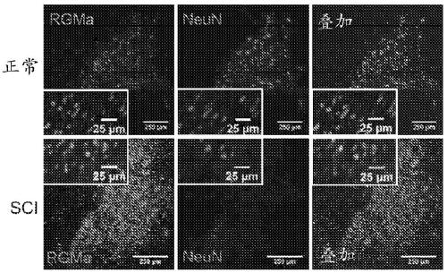
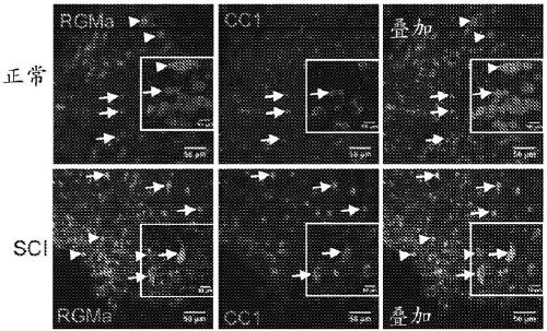
[0180] RGMa expression in rat and human spinal cord after SCI
[0181] Methodology: Rat tissue sections were prepared for immunohistochemical staining and incubated overnight at 4°C with primary antibodies. The following primary antibodies were used: NeuN for neurons (1:500, Millipore Bioscience Research Reagents), GFAP for astrocytes (1:200, Millipore), activated giant Iba-1 for phagocytes / microglia (1:1000, and Wako Chemicals), CS56 for chondroitin sulfate proteoglycan (1:500, Sigma), calcitonin for sensory fibers CGRP (1:1000, Millipore), 5HT for serotonergic fibers (1:3000, ImmunoStar), and hIgG for detecting human IgG antibodies (1:500, Millipore Inc. (Millipore). Sections were incubated overnight with primary antibodies diluted in blocking solution, washed, and incubated with fluorescently-conjugated secondary antibodies.
[0182] Human tissue sections were prepared for staining and incubated overnight with primary antibodies (1 :200 RGMa, Abcam; or 1 :100 Regenerate, ...
example 12
[0186] Effect of anti-RGMa antibody on neurite outgrowth in vitro
[0187] Methodology: For neurite outgrowth assays, E18 mouse cortical neurons were plated on poly-L-lysine treated with laminin (Invitrogen; 10 mg / ml) and RGMa protein (5 mg / ml). Amino acid-coated glass coverslips and incubated with control antibody (hIgG) or anti-RGMa (1 mg / ml) for 24 hours at 37°C. For neurite outgrowth analysis, cortical cells were immunostained with βIII-tubulin (Sigma; 1:500). For western blotting, mouse cortical neurons were lysed in RIPA buffer and loaded onto 10% acrylamide gels, then transferred to nitrocellulose membranes. Blots were probed with anti-RGMa antibodies (AE12-1 and AE12-1-Y) and anti-regenerate protein (E20; Santa Cruz; 10 mg / ml).
[0188] Results: In Western blot of mouse cortical neuron lysates, both AE12-1 and AE12-1-Y specifically detected a 50kDa band ( Figure 3A ). Cultured mouse primary cortical neurons were also shown to express RGMa, as shown by immunostaini...
example 13
[0190] Detection of anti-RGMa antibodies in serum, CSF, and spinal cord
[0191] Methodology: Cerebrospinal fluid (CSF) was sampled via lumbar puncture (LP) 6 weeks after SCI. Nine weeks after SCI, 3 weeks after the last antibody administration, serum was collected and rats were perfused. Antibody concentrations in CSF and serum samples from AE12-1, AE12-1-Y, and hlgG-treated rats were determined using ELISA assays.
[0192] Results: Antibody concentrations in the CSF of rats injected with AE12-1 ranged from 0.25-8.20 μg / ml, and for AE12-1-Y, antibody concentrations ranged from 0.33-6.77 μg / ml ( Figure 4B ). In contrast, antibody concentrations were considerably higher in serum, approximately 10-fold greater than in CSF, as antibodies were injected intravenously after the initial intraspinal injection immediately after SCI ( Figure 4C ). In addition, antibody concentrations continued to rise in serum 3 weeks after the last dose. Human antibodies were detected in the inj...
PUM
 Abstract
Description
Claims
Application Information
 - R&D Engineer
- R&D Manager
- IP Professional
- Industry Leading Data Capabilities
- Powerful AI technology
- Patent DNA Extraction
Browse by: Latest US Patents, China's latest patents, Technical Efficacy Thesaurus, Application Domain, Technology Topic, Popular Technical Reports.
© 2024 PatSnap. All rights reserved.Legal|Privacy policy|Modern Slavery Act Transparency Statement|Sitemap|About US| Contact US: help@patsnap.com