A device and method for segmenting cervical image lesion regions based on classification prior

A lesion area and image technology, applied in the field of cervical image lesion area segmentation device based on classification prior, can solve the problems of inability for doctors to locate biopsy lesions, strong subjectivity, and high professional requirements

Active Publication Date: 2022-04-01
SHANDONG IND TECH RES INST OF ZHEJIANG UNIV
View PDF3 Cites 0 Cited by
  • Summary
  • Abstract
  • Description
  • Claims
  • Application Information

AI Technical Summary

Problems solved by technology

Although colposcopy combined with biopsy can increase the positive detection rate, the location of biopsy lesions still requires a lot of medical experience of doctors
And there are two main problems in colposcopy examination: (1) High professional requirements, colposcopy images contain complex features such as texture and color, and the analysis of cervical tissue morphology under colposcopy requires a high degree of professional knowledge, so this examination requires rich experience (2) Strong subjectivity, the diagnosis of cervical images under colposcopy contains the subjective opinions of gynecologists, and there are more or less differences among experts in the diagnosis of some controversial images
At present, many machine learning and image processing methods have been applied to the field of colposcopy detection assistance, including the detection of cervical os, the detection of vinegar white area, etc. These methods indirectly assist doctors to make more accurate judgments to a certain extent. However, it cannot directly help doctors locate the location of biopsy lesions

Method used


Image

  • A device and method for segmenting cervical image lesion regions based on classification prior
  • A device and method for segmenting cervical image lesion regions based on classification prior
  • A device and method for segmenting cervical image lesion regions based on classification prior

Examples


Embodiment Construction

[0038] The present invention will be further described in detail below with reference to the accompanying drawings and embodiments. It should be noted that the following embodiments are intended to facilitate the understanding of the present invention, but do not limit it in any way.

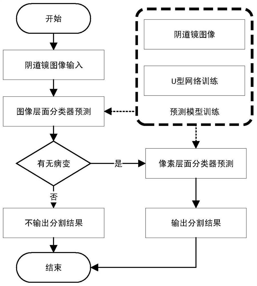
[0039] The cervical image lesion area segmentation device of the present invention includes:

[0040] An image acquisition device, used for acquiring images of the cervix treated with 3%-5% acetic acid solution;

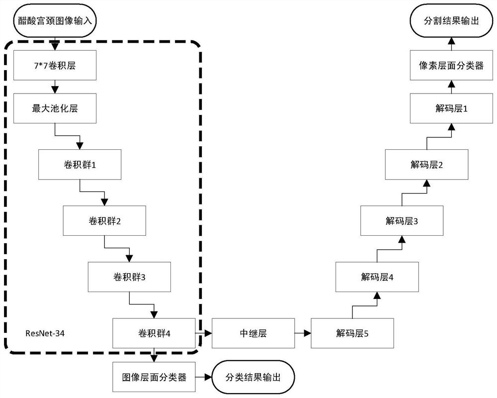
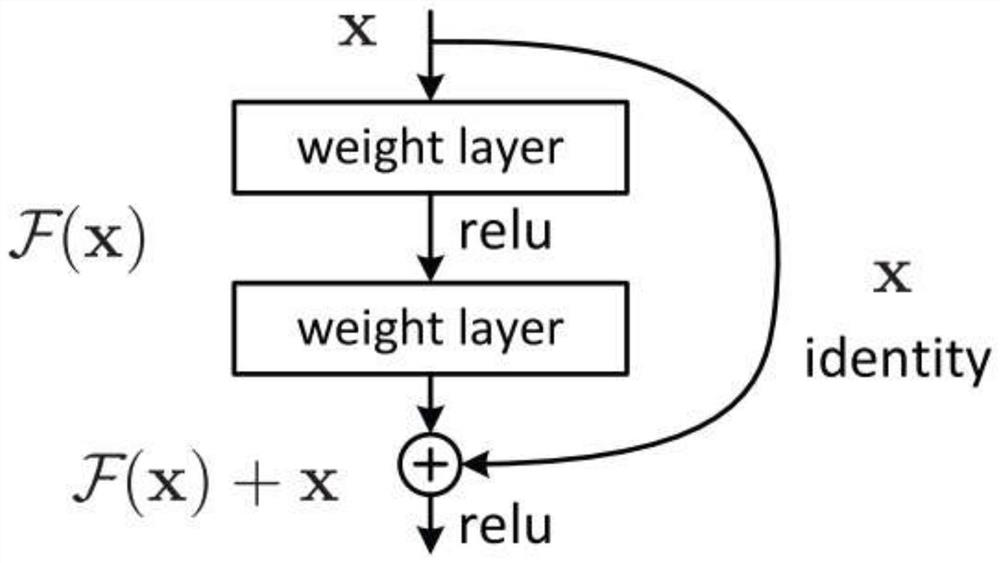
[0041] The processor includes a cervical image preprocessing module and a processing module, and the processing module includes a segmentation network model composed of a U-shaped network based on a residual connection, an image-level classifier and a pixel-level classifier, for outputting predictions on the cervical image. The location of the lesion area;

[0042] A memory for storing parameters of the segmentation network model in the processor;

[0043] The display device is used fo...


PUM


Abstract

The invention discloses a device and method for segmenting cervical image lesion regions based on classification prior, wherein the device includes: an image acquisition device for collecting cervical images processed by 3%-5% acetic acid solution; a processor including cervical An image preprocessing module and a processing module, the processing module including a segmentation network model composed of a U-shaped network based on a residual connection, an image level classifier and a pixel level classifier, for outputting and predicting the position of the lesion area on the cervical image; The memory is used for storing the parameters of the segmentation network model in the processor; the display device is used for displaying the predicted lesion area output by the processor. The present invention can provide doctors with high-accuracy biopsy lesion position candidate regions, and improve the detection rate of biopsy positives.

Description

technical field [0001] The invention belongs to the field of medical image processing, and in particular relates to a device and method for segmenting cervical image lesion regions based on classification prior. Background technique [0002] Cervical cancer, also known as cervical cancer, is cancer that occurs in the cervix. Worldwide, cervical cancer is the fourth most common cancer and the fourth most common cause of cancer death in women. However, from a normal cervix to HPV infection, a very small number of cervical lesions will further develop into cervical cancer, and this process generally takes about 5-10 years. Therefore, if certain detection techniques can be used to detect abnormalities in the cervix at the stage of cervical precancerous lesions, then there will be a great possibility to prevent cervical cancer. Cervical high-grade squamous intraepithelial lesion (High-grade squamous intraepithelial lesion, HSIL) is the last process of cervical cancer formation,...

Claims


Application Information

Patent Timeline
Patent Type & Authority Patents(China)
IPC IPC(8): G06T7/11G06V10/764G06V10/762G06V10/774G06K9/62A61B1/303A61B1/00
CPCA61B1/00009A61B1/303G06T7/11G06T2207/20081G06T2207/30096G06F18/23G06F18/24G06F18/214
Inventor 吴健马鑫军陈婷婷刘雪晨雷璧闻王文哲陆逸飞吕卫国袁春女姚晔俪王新宇吴福理
Owner SHANDONG IND TECH RES INST OF ZHEJIANG UNIV
Why Eureka
Try Eureka