
Blood vessel segmentation method and device, electronic device and storage medium

A blood vessel and segmentation technology, which is applied in the field of medical image processing, can solve the problems of limited blood vessel segmentation accuracy, few blood vessel branches, and blocky segmentation, and achieve the effect of improving the accuracy of blood vessel segmentation

Inactive Publication Date: 2019-04-23
SHANGHAI UNITED IMAGING INTELLIGENT MEDICAL TECH CO LTD
View PDF3 Cites 17 Cited by
  • Summary
  • Abstract
  • Description
  • Claims
  • Application Information

AI Technical Summary

Problems solved by technology

[0003] At present, a common method of blood vessel segmentation is traditional image processing methods, such as threshold segmentation method or region growing method, etc., but the accuracy of blood vessel segmentation of these methods is limited, and it is difficult to meet clinical needs.
Another commonly used method is the image processing method based on deep learning, such as fully convolutional neural network model (Fully convolutional networks, FCN), the fully convolutional neural network model U-net based on two-dimensional medical images, and the fully convolutional neural network model based on three-dimensional medical images. Convolutional neural network model V-net, etc. These methods are far superior to traditional image processing methods in terms of accuracy and robustness, but due to the limitations of imaging equipment and the complexity of the morphological structure of blood vessels, using any of the above convolution The blood vessel segmentation results obtained by the neural network model are prone to problems such as fewer blood vessel branches, wrong blood vessel borders, and segmented blocks.

Method used


Image

  • Blood vessel segmentation method and device, electronic device and storage medium
  • Blood vessel segmentation method and device, electronic device and storage medium
  • Blood vessel segmentation method and device, electronic device and storage medium

Examples


Embodiment 1

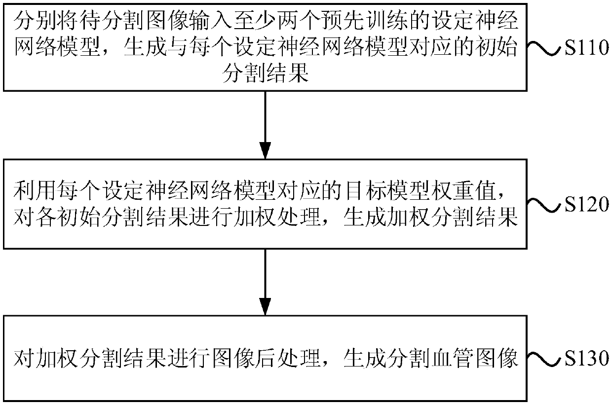
[0026] The blood vessel segmentation method provided in this embodiment is applicable to blood vessel segmentation of medical images, especially for blood vessel segmentation in complex organs, such as liver blood vessel segmentation. The method can be performed by a blood vessel segmentation device, which can be realized by software and / or hardware, and which can be integrated in electronic equipment with image processing functions, such as desktop computers or servers. see figure 1 , the method of this embodiment specifically includes the following steps:

[0027] S110. Input the image to be segmented into at least two pre-trained preset neural network models, and generate an initial segmentation result corresponding to each preset neural network model.

[0028] Wherein, the image to be segmented refers to a medical image including blood vessels to be segmented, which may be a two-dimensional medical image or a three-dimensional medical image. Setting the neural network mo...

Embodiment 2

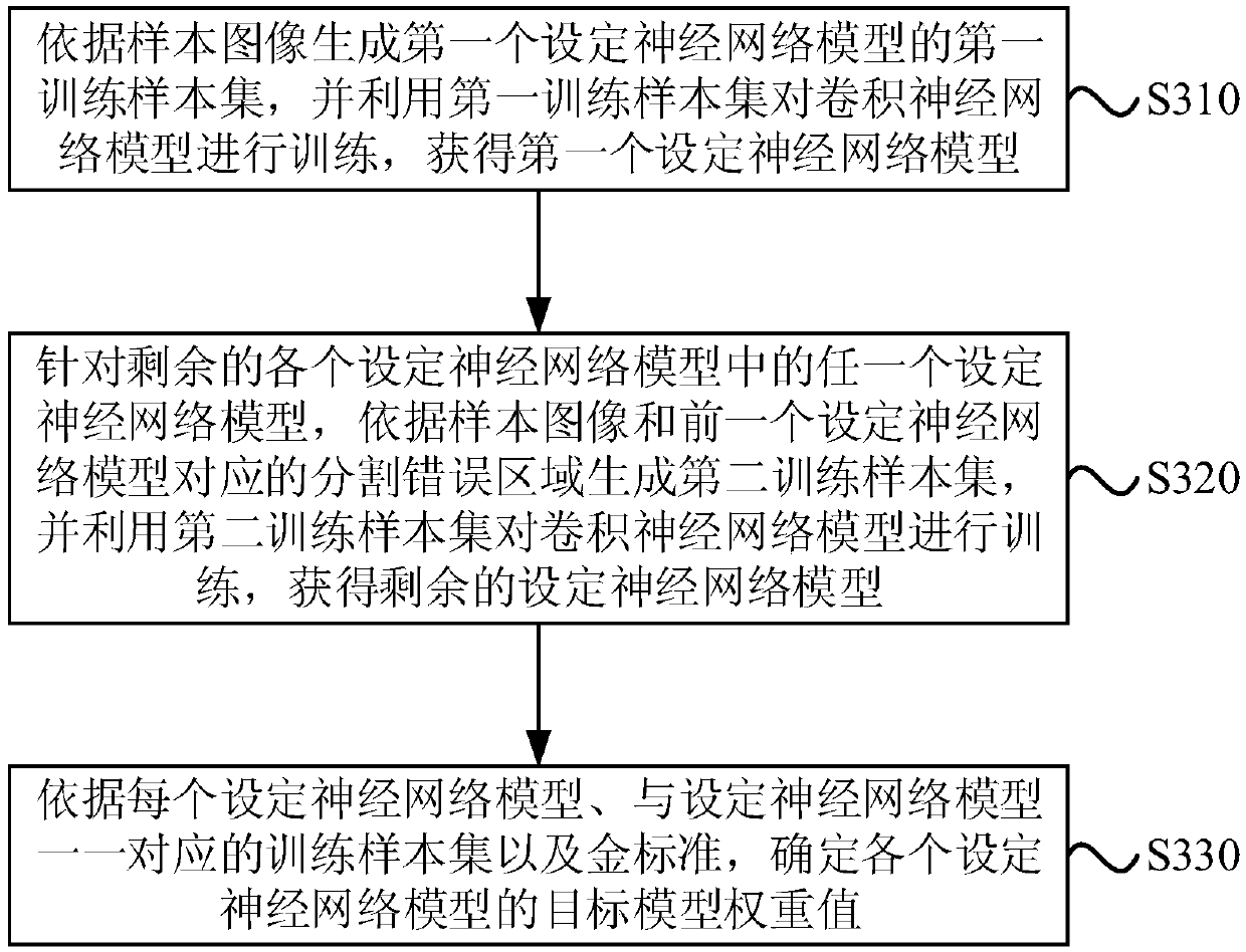
[0045] Based on the above-mentioned embodiments, this embodiment describes the training method of the blood vessel segmentation model. The explanations of terms that are the same as or corresponding to the above-mentioned embodiments will not be repeated here. see image 3 , the model training method for blood vessel segmentation provided in this embodiment includes:

[0046] S310. Generate a first training sample set of a first set neural network model according to the sample image, and use the first training sample set to train the convolutional neural network model to obtain a first set neural network model.

[0047] Among them, the sample image refers to the medical image containing blood vessels used for the training of the blood vessel segmentation model. In order to improve the applicability of the blood vessel segmentation model, medical images of various organs can be selected, such as brain medical images, liver medical images and limbs medical images, etc. The fi...

Embodiment 3

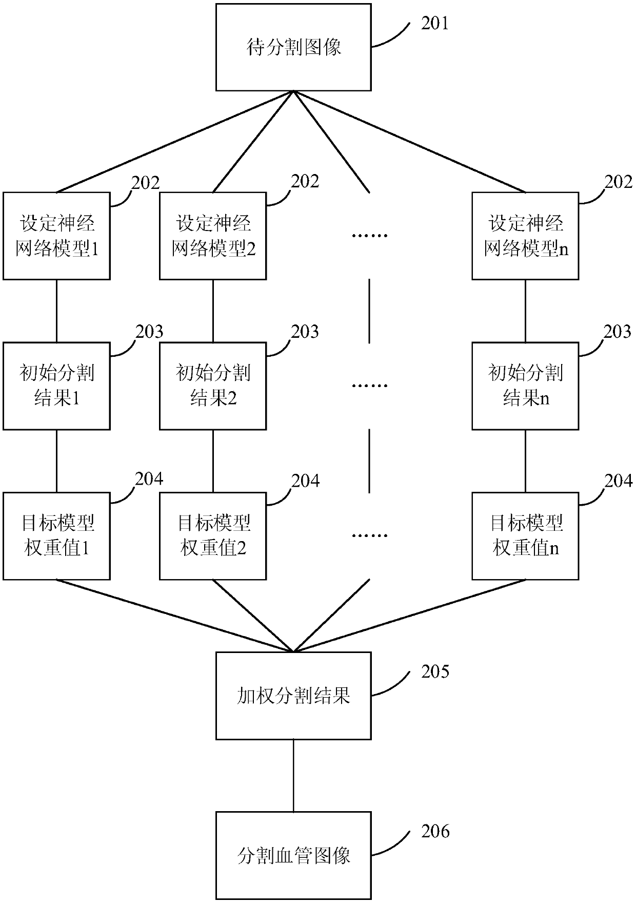
[0071] This embodiment provides a blood vessel segmentation device, see Figure 4 , the device specifically includes:

[0072] The initial segmentation result generation module 410 is used to input the image to be segmented into at least two pre-trained neural network models to generate at least two initial segmentation results;

[0073] The weighted segmentation result generation module 420 is used to use the target model weight value corresponding to each set neural network model to carry out weighted processing on each initial segmentation result to generate a weighted segmentation result, wherein the target model weight value is higher than that of the training set neural network. Determined when the model;

[0074] The segmented blood vessel image generation module 430 is configured to perform image post-processing on the weighted segmentation result to generate a segmented blood vessel image.

[0075] Optionally, the initial segmentation result generating module 410 is...


PUM


Abstract

The embodiment of the invention discloses a blood vessel segmentation method and device, an electronic device and a storage medium. The method comprises the following steps of respectively inputting an image to be segmented into at least two pre-trained set neural network models, and generating an initial segmentation result corresponding to each set neural network model; carrying out weighting processing on each initial segmentation result by utilizing a target model weight value corresponding to each set neural network model to generate a weighted segmentation result, the target model weightvalue being determined when the set neural network model is trained; and carrying out image post-processing on the weighted segmentation result to generate a segmented blood vessel image. According to the technical scheme, the Boosting modeling idea is applied to the field of blood vessel segmentation for the first time, the problem that a blood vessel segmentation result obtained through a single neural network model is low in precision is solved, and the effect of improving the blood vessel segmentation precision is achieved.

Description

technical field [0001] Embodiments of the present invention relate to medical image processing technologies, and in particular to a blood vessel segmentation method, device, electronic equipment, and storage medium. Background technique [0002] Vessel segmentation in medical images is a fundamental problem. For example, the segmentation of blood vessels in the liver is widely used in the diagnosis and treatment of liver lesions and the planning of liver surgery. [0003] At present, a common method for blood vessel segmentation is traditional image processing methods, such as threshold segmentation method or region growing method, etc., but the accuracy of blood vessel segmentation of these methods is limited, and it is difficult to meet clinical needs. Another commonly used method is the image processing method based on deep learning, such as fully convolutional neural network model (Fully convolutional networks, FCN), the fully convolutional neural network model U-net bas...

Claims


Application Information

Patent Timeline
IPC IPC(8): G06T7/00G06T7/11
CPCG06T7/0012G06T7/11G06T2207/20084G06T2207/30101
Inventor 杨燕平高耀宗
Owner SHANGHAI UNITED IMAGING INTELLIGENT MEDICAL TECH CO LTD
Learn More