A biosensor for detecting uracil-DNA glycosylase and its preparation method

A biosensor and glycosylase technology, applied in the field of biosensors, can solve the problems of difficulty in generalization, complexity, time-consuming sensitivity, etc., and achieve the effects of reducing complexity, fast reaction speed, and sensitive detection

Active Publication Date: 2021-06-15
UNIV OF JINAN
View PDF10 Cites 0 Cited by
  • Summary
  • Abstract
  • Description
  • Claims
  • Application Information

AI Technical Summary

Problems solved by technology

[0003] Traditional UDG activity detection methods include gel electrophoresis, nucleic acid labeling and other methods, however, they require complex nucleic acid labeling and gel electrophoresis procedures, are time-consuming and have poor sensitivity, and are difficult to generalize
In order to overcome the above defects, some UDG activity detection methods based on colorimetry and fluorescence have been developed. These new technologies have brought great progress to the detection of UDG activity; however, further improvement is needed to achieve more sensitive and specific detection of UDG

Method used


Image

  • A biosensor for detecting uracil-DNA glycosylase and its preparation method
  • A biosensor for detecting uracil-DNA glycosylase and its preparation method
  • A biosensor for detecting uracil-DNA glycosylase and its preparation method

Examples


Embodiment 1

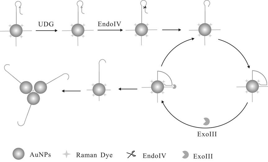
[0045] The steps for modifying Hairpin Probe and Track DNA to the surface of gold nanoparticles are as follows:

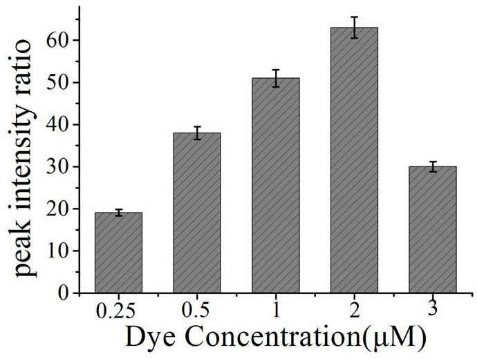
[0046] a. Take 1 mL nano-gold solution in a centrifuge tube, centrifuge for 15 min, and centrifuge the two tubes at the same time for later use. Centrifuge until the supernatant is colorless and transparent, remove the supernatant, and add 300 μL of sterile water to concentrate the nano-gold solution to 1 nM. Transfer to a 1 mL glass bottle, seal it with tin foil, and add different volumes of Raman dyes (4NTP final concentrations are 0.25uM, 0.5 uM, 1uM, 2 uM and 3 uM, respectively).

[0047] b. After standing at room temperature for 30 min, add 150 μL of -SH-modified substrate probe (Hairpin Probe and Track DNA) with a concentration of 10 μM, mix well, and place at 4 °C for 24 h.

[0048] c. Slowly add 50 μL of PBS buffer several times, add magnets (soaked in aqua regia the day before) and stir for 10 min, then continue to add 27 μL of PBS buffer. Take out the m...

Embodiment 2

[0055] The steps for modifying Hairpin Probe and Track DNA to the surface of gold nanoparticles are as follows:

[0056] a. Take 1 mL nano-gold solution in a centrifuge tube, centrifuge for 15 min, and centrifuge the two tubes at the same time for later use. Centrifuge until the supernatant is colorless and transparent, remove the supernatant, and add 300 μL of sterile water to concentrate the nano-gold solution to 1 nM. Transfer to a 1 mL glass bottle, seal it with tin foil, and add Raman dye (4NTP final concentration is 2 μM).

[0057] b. After standing at room temperature for 30 min, add 150 μL of -SH-modified substrate probe (Hairpin Probe and Track DNA) with a concentration of 10 μM, mix well, and place at 4 °C for 24 h.

[0058] c. Slowly add 50 μL of PBS buffer several times, add magnets (soaked in aqua regia the day before) and stir for 10 min, then continue to add 27 μL of PBS buffer. Take out the magnet, and place it at 4°C for 48 h.

[0059] d. After 48 hours, sl...

Embodiment 3

[0066] The steps for modifying Hairpin Probe and Track DNA to the surface of gold nanoparticles are as follows:

[0067] a. Take 1 mL nano-gold solution in a centrifuge tube, centrifuge for 15 min, and centrifuge the two tubes at the same time for later use. Centrifuge until the supernatant is colorless and transparent, remove the supernatant, and add 300 μL of sterile water to concentrate the nano-gold solution to 1 nM. Transfer to a 1 mL glass bottle, seal it with tin foil, and add Raman dye (4NTP final concentration is 2 μM).

[0068] b. After standing at room temperature for 30 min, add 150 μL of -SH-modified substrate probe (Hairpin Probe and Track DNA) with a concentration of 10 μM, mix well, and place at 4 °C for 24 h.

[0069] c. Slowly add 50 μL of PBS buffer several times, add magnets (soaked in aqua regia the day before) and stir for 10 min, then continue to add 27 μL of PBS buffer. Take out the magnet, and place it at 4°C for 48 h.

[0070] d. After 48 hours, sl...


PUM


Abstract

The invention relates to the technical field of biosensors, in particular to biosensors based on nano-gold DNA molecular machines and surface-enhanced Raman scattering. In order to solve the problems of relatively low specificity and sensitivity and high cost of methods for detecting UDG activity in the prior art. A biosensor based on surface-enhanced Raman scattering of DNA molecular machines to detect UDG activity, combining nano-gold molecular machines with surface-enhanced Raman, and a homogeneous reaction mixture. Preparation method: synthesize gold nanoparticles; modify Hairpin Probe, Track DNA and Raman dyes on the surface of gold nanoparticles; mix labeled nano gold solution with homogeneous reaction solution. The specific cleavage of endonuclease IV is used to realize the opening of the DNA molecular machine, and the surface-enhanced Raman detection is used to realize high-sensitivity detection; the use of exonuclease III realizes the circulation of the DNA molecular machine and plays the role of signal amplification.

Description

technical field [0001] The invention belongs to the technical field of biosensors, in particular to a biosensor for detecting uracil-DNA glycosylase activity based on surface-enhanced Raman scattering of DNA molecular machines, and also relates to a preparation method thereof. Background technique [0002] The genome consists of specific pairings of DNA bases, the stability and accuracy of which are prerequisites for all living organisms. However, the structural properties of DNA bases can be disrupted by various environmental factors and endogenous reactive oxygen species, leading to genome instability and induction of carcinogenesis. Uracil is a common damaged base in DNA. During DNA replication, dUTP is misincorporated or cytosine is hydrolyzed and deaminated, resulting in the conversion of G:C base pairs into A:U base pairs, which eventually leads to gene mutation. Uracil in DNA is usually repaired by uracil-DNA glycosylase (UDG) in the base excision repair system. Ur...

Claims


Application Information

Patent Timeline
Patent Type & Authority Patents(China)
IPC IPC(8): G01N21/65
Inventor 王玉李莎莎刘素黄加栋张儒峰赵一菡瞿晓南孙文玉王业茹张曼如
Owner UNIV OF JINAN
Why Eureka
Try Eureka