

A semi-automatic segmentation sequential optimization registration method
A sequential optimization, semi-automatic technology, applied in image analysis, image enhancement, instrument and other directions, can solve the problems of increased number of registrations, time-consuming, registration errors, etc., and achieve the effect of reducing time-consuming
- Summary
- Abstract
- Description
- Claims
- Application Information
AI Technical Summary
Problems solved by technology
Method used
Image

Examples
Embodiment Construction
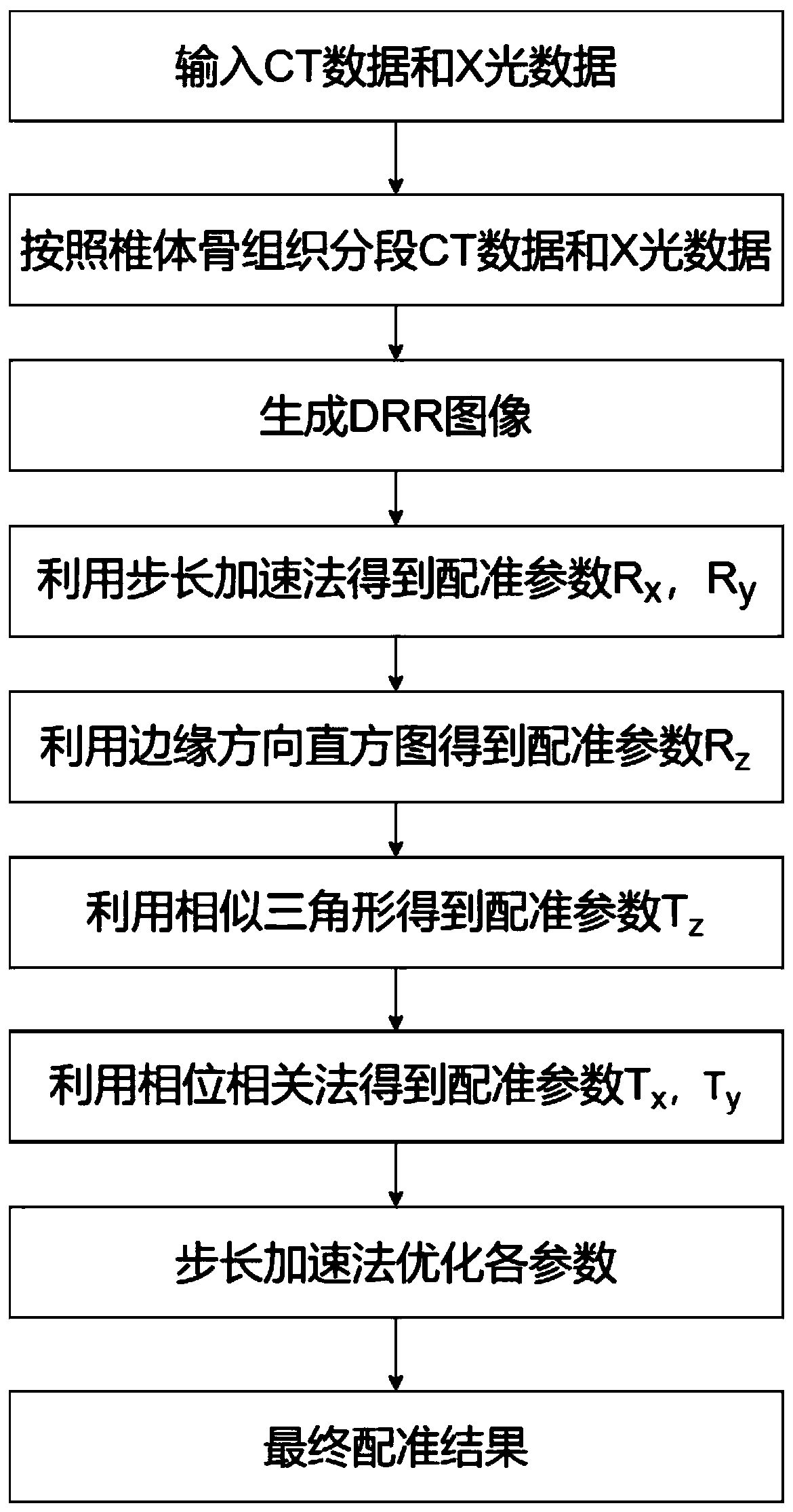
[0033] The technical solutions of the present invention will be described more clearly and completely below in conjunction with the accompanying drawings and the embodiments of the present invention.
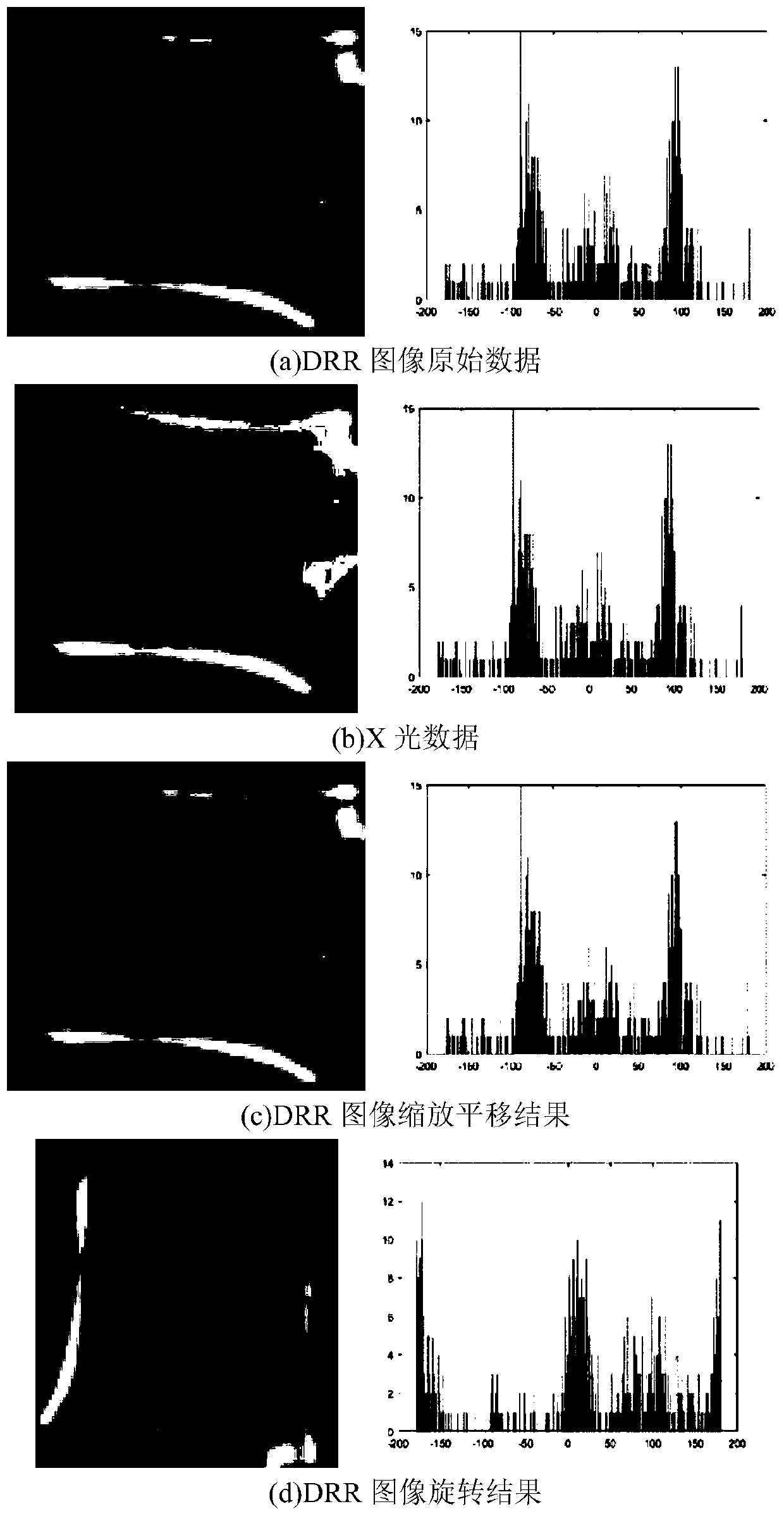
[0034] In this embodiment, CT images and X-ray images of the spine of the Third Hospital of Peking University were used to intercept the CT and X-ray images of the lesion, including three segments of bone tissue in total. The CT image volume data size is 512×359×238, and the slice thickness is 0.7mm. X-ray image size is 1024×803. The resolution is 0.28346mm / pixel.
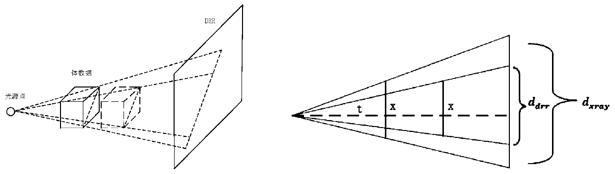
[0035] The volume data described in this method is a term in three-dimensional graphics visualization, and the volume data is composed of voxels. Voxel is the basic volume element, and can also be understood as a point or a small area with arrangement and color in three-dimensional space. The CT image volume data used in this embodiment is similar to cuboid data in which all slices are superimposed without segment...
PUM
 Abstract
Description
Claims
Application Information