Nucleic acid aptamer fluorescence sensor for detecting myoglobin and construction method of nucleic acid aptamer fluorescence sensor

A nucleic acid aptamer and fluorescent sensor technology, which is applied in the field of analysis and detection, can solve the problems of cumbersome time-consuming, cumbersome separation and washing steps, and high cost, and achieve the effects of improving accuracy and sensitivity, shortening fluorescence recovery time, and fewer detection steps

Inactive Publication Date: 2019-06-18
JIAXING UNIV
View PDF0 Cites 6 Cited by
  • Summary
  • Abstract
  • Description
  • Claims
  • Application Information

AI Technical Summary

Problems solved by technology

[0005] At present, the methods for detecting myoglobin mainly include liquid chromatography, mass spectrometry, surface-enhanced Raman scattering method, giant magnetic impedance immunoassay, electrochemical immunoassay, fluorescence method, surface plasmon resonance method, and enzyme-linked immunosorbent assay. ELISA is a classic and widely used clinical immunoassay method, which is suitable for batch specimen detection, but this method has many influencing factors and separation And the washing steps are cumbersome, the measurement cycle is too long, etc.
Although the chemiluminescence method is safe, sensitive, and versatile, the instrument cost of this method is relatively high
Although mass spectrometry has high sensitivity and specificity, the instrument is relatively expensive, and its database is not perfect at present, so it has not been widely used.
In addition, most of the above-mentioned methods for detecting myoglobin are detection systems based on the mutual recognition of antibody antigens. Its advantage is that immunoassay has high selectivity and sensitivity, but the preparation of antibodies requires immunization animal experiments or cell experiments, which is cumbersome and time-consuming. ,high cost
Moreover, antibodies are easily affected by external conditions, especially temperature, and require harsh storage conditions, which largely limits the flexible application of these methods

Method used


Image

  • Nucleic acid aptamer fluorescence sensor for detecting myoglobin and construction method of nucleic acid aptamer fluorescence sensor
  • Nucleic acid aptamer fluorescence sensor for detecting myoglobin and construction method of nucleic acid aptamer fluorescence sensor
  • Nucleic acid aptamer fluorescence sensor for detecting myoglobin and construction method of nucleic acid aptamer fluorescence sensor

Examples


Embodiment 1

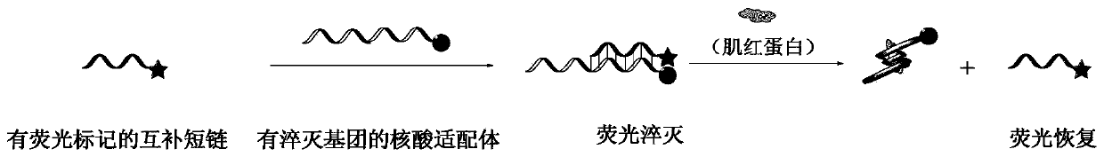
[0042] 1. The base series structure of the nucleic acid aptamer with a quenching group is:

[0043] 5'-CCCTCCTTTCCTTCGACGTAGATCTGCTGCGTTGTTCCGA-

[0044] DABCYL-3', wherein DABCYL is a quenching group, and its molecular structure is: 4-[4-(dimethylamino)phenylazo]benzoic acid.

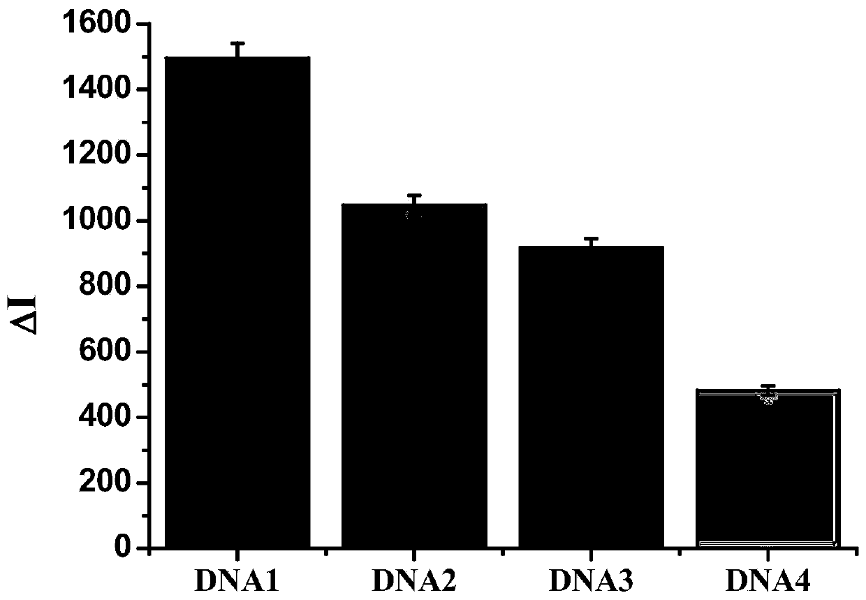
[0045] The base series structure of the complementary short chain with a fluorescent group is: DNA1: 5'-FAM-GGAACAACGC-3'; DNA2: 5'-FAM-TCGGAACAAC-3'; DNA3: 5'-FAM-CGGAACAACG-3 '; DNA4: 5'-FAM-GAACAACGCA-3', wherein FAM is a fluorescent group, and its molecular structure is: 6-carboxyfluorescein.

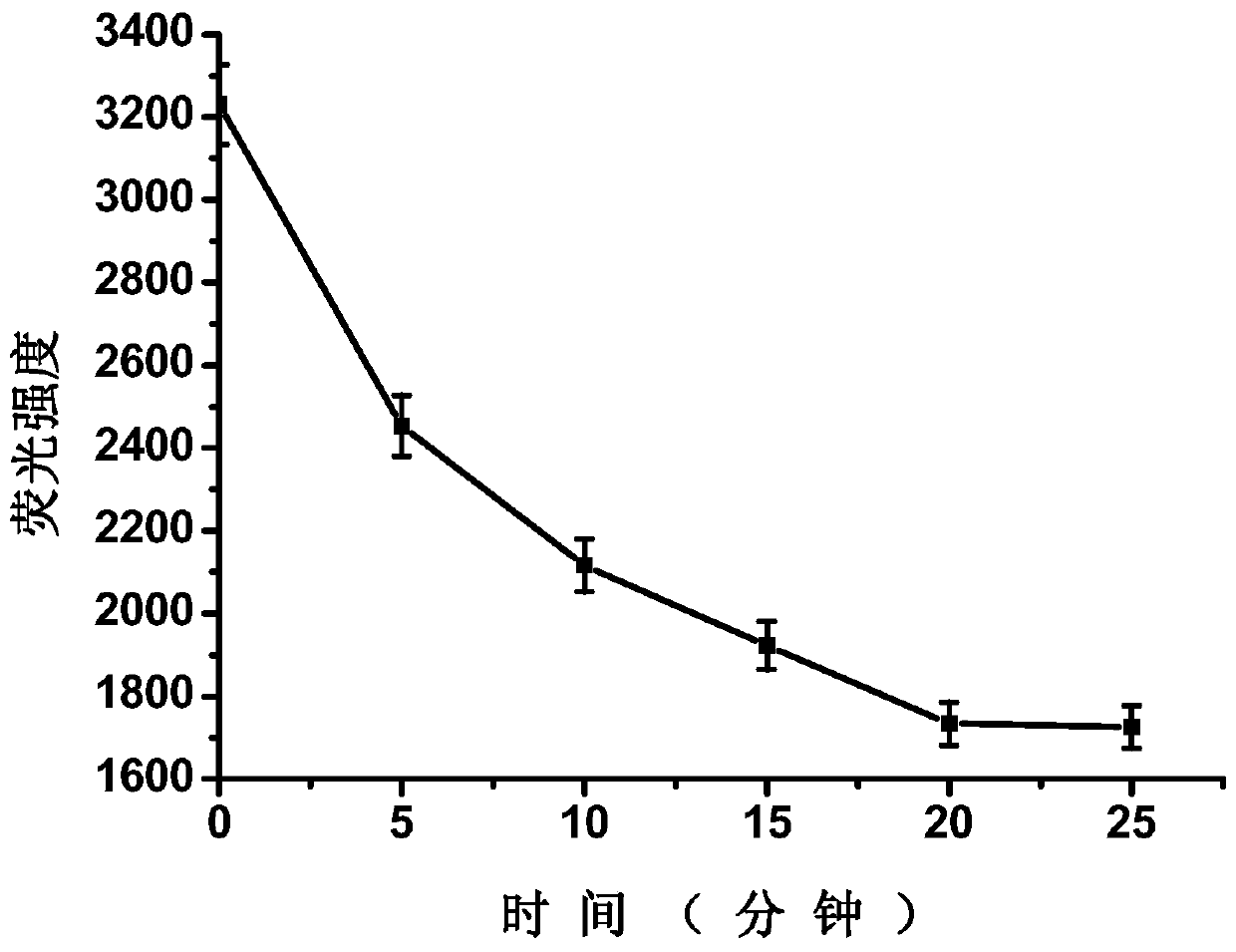
[0046] 2. Add 1760 μL of 0.01M PBS buffer solution (pH=7.4), 100 μL of 500 nM complementary short chain with fluorescent group and 100 μL of 500 nM nucleic acid aptamer with quencher group into a 5 mL plastic tube in sequence. The final concentrations of the complementary short chains linked with fluorescent groups and the nucleic acid aptamers linked with quenching groups were both 25 nM. The mixed solut...

Embodiment 2

[0060] Example 2: Detection of human serum myoglobin samples and spiked samples

[0061] 1. First, the human serum sample (33.4ng / mL, the test result of the hospital) was diluted 6.68 times with 0.01M PBS buffer solution (pH=7.4), and then the macromolecules in the serum were filtered out with an ultrafiltration centrifuge tube (30kD) Protein, human serum myoglobin samples can be prepared.

[0062] Next, add 1760 μL of 0.01M PBS buffer solution (pH=7.4), 100 μL of 500 nM complementary short chains with fluorescent groups and 100 μL of 500 nM nucleic acid aptamers with quenching groups into a 5 mL plastic tube in sequence. The final concentrations of the complementary short chains linked with fluorescent groups and the nucleic acid aptamers linked with quenching groups were both 25 nM. The mixed solution was incubated at 25°C for 20 minutes.

[0063] Then, 1960 μL of the mixed solution was divided into two equal parts, each solution was 980 μL, and 20 μL of 0.01M PBS buffer s...


PUM

PropertyMeasurementUnit
wavelengthaaaaaaaaaa

Abstract

The invention discloses a nucleic acid aptamer fluorescence sensor for detecting myoglobin and a construction method of the nucleic acid aptamer fluorescence sensor and belongs to the technical fieldof analysis and detection. The construction method includes the following steps that: a myoglobin nucleic acid aptamer modified with a quencher and a nucleic acid aptamer complementary strand modifiedwith a fluorophore are added into a buffer system so as to be hybridized, and therefore, a nucleic acid aptamer fluorescence sensor can be prepared; when myoglobin is added, the nucleic acid aptameris separated from the complementary short strand, and is combined with the myoglobin; the complementary short strand connected with the fluorophore is separated from the nucleic acid aptamer connectedwhich the quencher, so that the fluorescence of the fluorophore is recovered; and the intensity of the recovery of the fluorescence is proportional to the concentration of the myoglobin, and therefore, the concentration of the myoglobin can be measured. According to the sensor of the invention, the nucleic acid aptamer fluorescence sensor which is inexpensive and easy to prepare is adopted to detect the myoglobin, and therefore, the myoglobin can be accurately detected. The method has the advantages of simple design, low cost, few detection steps, simple operation, high sensitivity and good selectivity.

Description

technical field [0001] The invention relates to the technical field of analysis and detection, in particular to a nucleic acid aptamer fluorescence sensor for detecting myoglobin and a construction method thereof. Background technique [0002] Myoglobin is a small protein that transports and stores oxygen in muscle cells. In normal human serum, the content of myoglobin is very small, about 30-90ng / mL. When the myocardium is damaged, myoglobin will be quickly released from the damaged cells into the blood, resulting in the loss of myoglobin in the blood. The content is significantly increased, and can rise to 200-900ng / mL, so it can be used as a marker for early diagnosis of acute myocardial infarction. [0003] Acute myocardial infarction is a common clinical cardiovascular disease. The disease is caused by myocardial ischemia and hypoxia, and has the characteristics of acute onset and high mortality, which seriously threatens the life safety of patients. The key to clini...

Claims


Application Information

Patent Timeline
Patent Type & Authority Applications(China)
IPC IPC(8): G01N33/68G01N21/64
CPCG01N21/6428G01N21/6486G01N33/6893G01N2021/6432
Inventor 杨义文李蕾刘东奎曾延波王海龙翟云云张剑路易霞
Owner JIAXING UNIV
Why Eureka
Try Eureka