miRNA detection fluorescent biological probe and detection method and application thereof

A fluorescent biological probe and fluorescence technology, which is applied in the field of biosensors, can solve the problem that miRNA detection methods cannot be fast, simple and low-cost at the same time, and achieve the effects of label-free, high specificity and simple steps.

Active Publication Date: 2019-06-21
JIANGSU INST OF NUCLEAR MEDICINE
View PDF3 Cites 12 Cited by
  • Summary
  • Abstract
  • Description
  • Claims
  • Application Information

AI Technical Summary

Benefits of technology

This patented inventive device uses two different reactions systems that work together effectively at room temperature without complicated procedures or requiring expensive equipment like laser scanning microscopes. These reactors are designed to have very small amounts of probes attached to them while still being able to measure their presence accurately. They also use special materials called quantum dots instead of gold colloids to improve sensitiveness over traditional methods such as enzyme linked immunosorbant assay techniques. Overall this innovation makes these devices highly sensitive but easy to operate even on tiny samples.

Problems solved by technology

This patented technical problem addressed by this patent relates to improving the accuracy and precision of identifying minute amounts of gene products called miR Nbspins from complex mixtures containing thousands or millions of genome elements without requiring expensive instrumentations like electronics. Current techniques require multiple step operations involving labeling and purifications beforehand, making them difficult to use clinically because they cannot provide real-time results during disease monitoring procedures.

Method used


Image

  • miRNA detection fluorescent biological probe and detection method and application thereof
  • miRNA detection fluorescent biological probe and detection method and application thereof
  • miRNA detection fluorescent biological probe and detection method and application thereof

Examples


Embodiment 1

[0077] Embodiment 1 detects the design of the fluorescent biological probe of miRNA

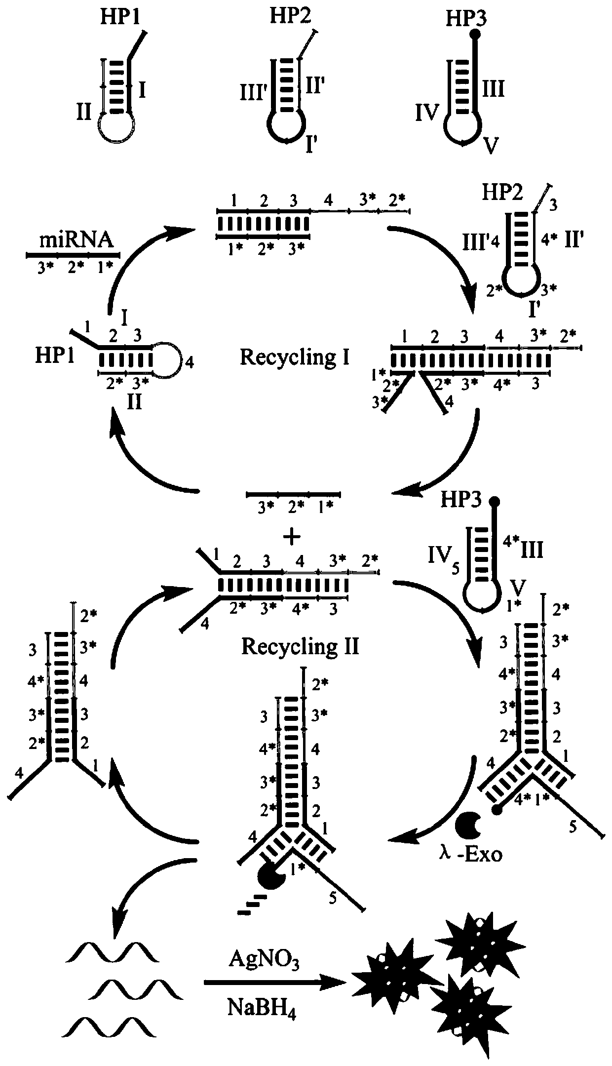
[0078] The fluorescent bioprobe for detecting miRNA in this embodiment includes a first hairpin, a second hairpin and a third hairpin; the first hairpin, the second hairpin and the third hairpin are all single-stranded linear molecules After self-folding, the complementary bases in the folding region are hybridized, and the part of the local region that forms a double-stranded structure is the stem region, and the part that does not form a double-stranded structure and folds back is the loop region;

[0079] The first hairpin includes: domain (I), which can hybridize complementary to the target miRNA; domain (II), which partially hybridizes with the domain (I) to form a hairpin structure;

[0080] The second hairpin includes: a structural domain (I'), which can partially hybridize with the structural domain (I) of the first hairpin; a structural domain (II'), which is connected to one end of ...

Embodiment 2

[0095] Embodiment 2 detects the fluorescent biosensor of miRNA

[0096] This embodiment provides a fluorescent biosensor for detecting miRNA, including an A reaction system and a B reaction system;

[0097] The A reaction system, in terms of 50 μL, includes:

[0098] miRNA to be tested, 4 μL;

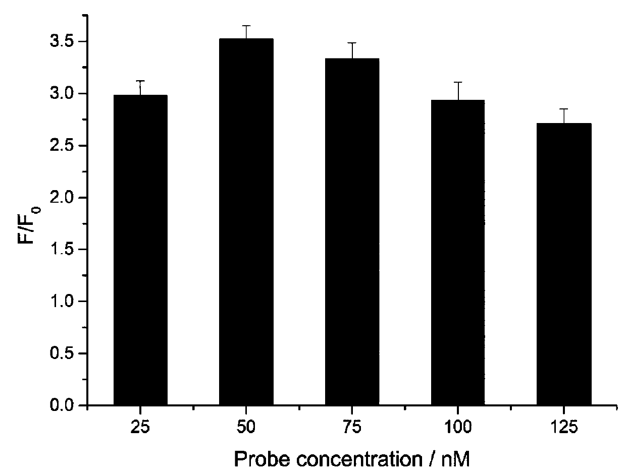
[0099] Probe solution, containing hairpin HP1, hairpin HP2 and hairpin HP3 in a molar ratio of 1:1:1, the concentration of hairpin HP1 is 50 nM (nmol / L), 17 μL;

[0100] The first buffer solution contains Tris-HNO with a concentration of 15mM 3 , a concentration of 55mM KNO 3 , a concentration of 5 mM Mg(NO 3 ) 2 , and DTT (dithiothreitol) at a concentration of 1.5 mM, pH 7.8, 18 μL;

[0101] The RNase inhibitor, 15U, 6 μL;

[0102] The exonuclease λ-Exo, 15U, 4 μL;

[0103] wxya 2 O make up.

[0104] The B reaction system, in terms of 50 μL, includes:

[0105] The second buffer containing Ag ions is AgNO 3 A sodium citrate solution in which AgNO 3 The molar ratio with the ...

Embodiment 3

[0108] Embodiment 3 detects the fluorescent biosensor of miRNA

[0109] This embodiment provides a fluorescent biosensor for detecting miRNA, including an A reaction system and a B reaction system;

[0110] The A reaction system, in terms of 50 μL, includes:

[0111] miRNA to be tested, 6 μL;

[0112] Probe solution, containing hairpin HP1, hairpin HP2 and hairpin HP3 in a molar ratio of 1:1:3, the concentration of hairpin HP1 is 50 nM (M is mol / L), 13 μL;

[0113] The first buffer solution contains Tris-HNO with a concentration of 25mM 3 , a concentration of 45mM KNO 3 , a concentration of 15 mM Mg(NO 3 ) 2 , and DTT (dithiothreitol) at a concentration of 0.5 mM, pH 8.0, 22 μL;

[0114] The RNase inhibitor, 5U, 4 μL;

[0115] The exonuclease λ-Exo, 25U, 6 μL;

[0116] Top up with deionized water.

[0117] The B reaction system, in terms of 50 μL, includes:

[0118] The second buffer containing Ag ions is AgNO 3 A sodium citrate solution in which AgNO 3 The molar r...


PUM


Abstract

The invention belongs to the technical field of biosensors and particularly relates to a miRNA detection fluorescent biological probe, a miRNA detection fluorescent sensor and application thereof anda miRNA detection method. In adoption of the probe for miRNA detection, steps are simple, marking is avoided, reaction is carried out in isothermal homogeneous solution, and problems of a complex separation process and high-time-consumption thermal cycle are avoided; a catalytic hairpin assembly technique and a lambda exonuclease (lambda-Exo) auxiliary signal amplification technique are ingeniously combined; by combination with DNA silver nano-clusters (DNA-AgNCs), high-sensitivity detection of biological targets is realized, the limit of detection reaches 0.89fM minimally, single base difference among miRNA family members can be distinguished, high specificity is realized, and the problem that a miRNA detection method in the prior art fails to achieve quickness, simplicity and low cost aswell as high detection sensitivity and high specificity.

Description


Claims


Application Information

Patent Timeline
Owner JIANGSU INST OF NUCLEAR MEDICINE
Why Eureka
Try Eureka