Coronary artery CT imaging method and computer readable storage medium
A coronary artery and CT imaging technology, applied in the field of medical imaging, can solve problems such as artifacts and cannot meet the needs of doctors for diagnosis
- Summary
- Abstract
- Description
- Claims
- Application Information
AI Technical Summary
Problems solved by technology
Method used
Image
Examples
Embodiment 1
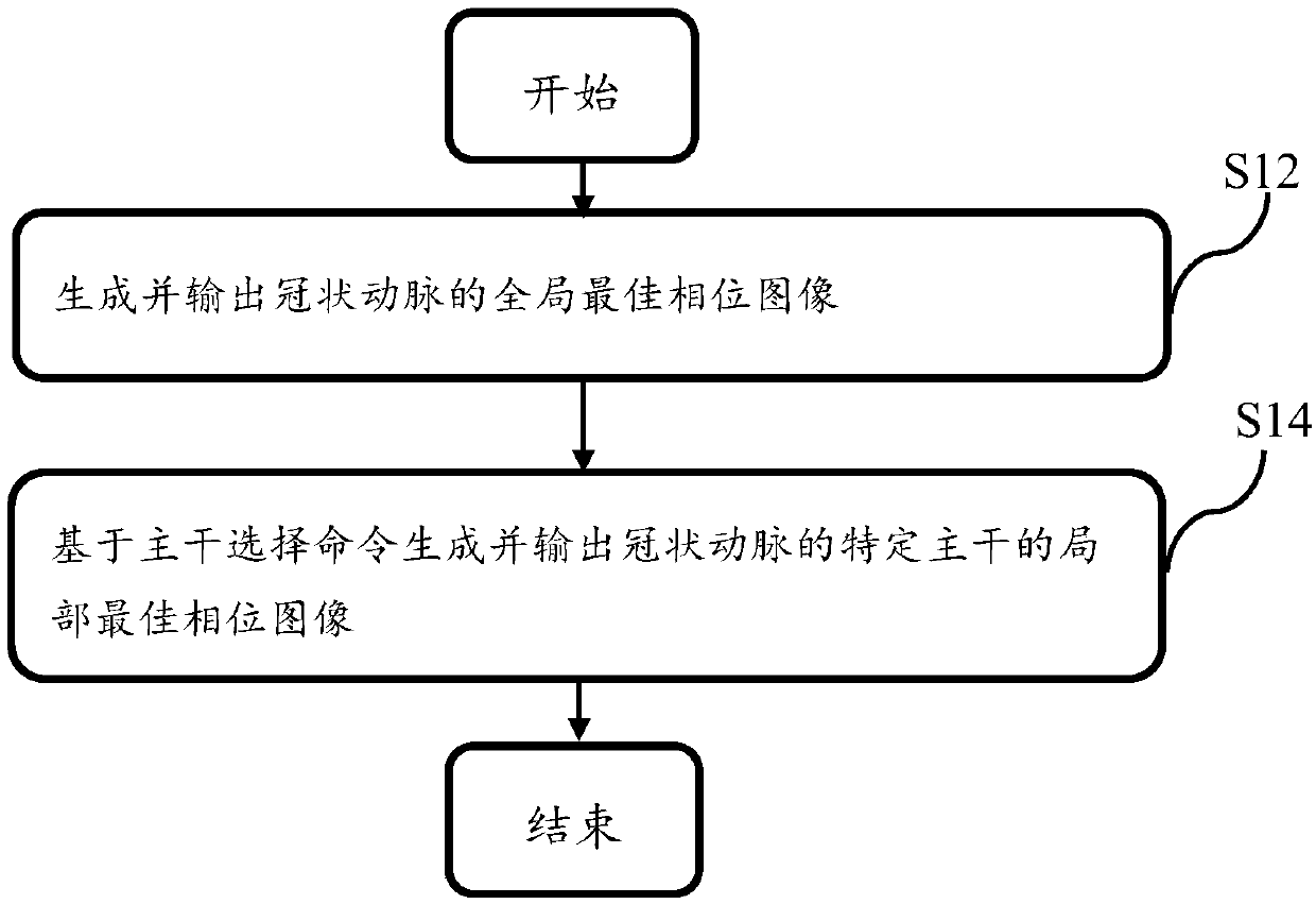
[0022] figure 1 Is a flowchart of the coronary artery CT imaging method according to the first embodiment of the present invention, such as figure 1 As shown, the method includes step S12 and step S14. In step S12, a global optimal phase image of the coronary artery is generated and output; in step S16, a local optimal phase image of a specific trunk of the coronary artery is generated and output based on the trunk selection command.
[0023] The aforementioned coronary arteries refer to arterial vessels located in the heart of the human body, and their main trunks may include the right coronary artery (RCA), the left anterior descending artery (LAD), and the left circumflex artery (LCX).
[0024] The above-mentioned global optimal phase image of the coronary arteries refers to the optimal phase of imaging for the entire coronary arteries. This phase may not be a better imaging phase for some of the main trunks; and the localization of the specific main trunk The best phase image r...
Embodiment 2
[0055] Figure 4 It is a flowchart of the coronary CT imaging method provided by the second embodiment of the present invention. Such as Figure 4 As shown, the method includes steps S41, S43, S45, and S47.
[0056] In step S41, image data of the coronary arteries are collected.
[0057] In step S43, the motion index of each main stem of the coronary artery at different phases is acquired based on the collected image data.
[0058] In step S45, the local optimal phase of each backbone is determined based on the motion index of each backbone at different phases.
[0059] In step S47, image reconstruction is performed based on the image data at the local optimal phase of the specific backbone to generate a local optimal phase image for the specific backbone.
[0060] After step S47, the generated local optimal phase image can be further output to the display device for display.
[0061] The content of each step of this embodiment has been described in the first embodiment, and will not ...
Embodiment 3
[0066] The embodiment of the present invention also provides a computer-readable storage medium for storing a computer program. When the computer program is installed in the computer system of the CT imaging system, it is used to perform the coronary CT imaging in the first embodiment above. method.
[0067] Specifically, the computer program can be used to make the computer system execute the following steps:
[0068] Step 1: Generate and output the global optimal phase image of the coronary artery;
[0069] Step 2: Generate and output a local optimal phase image of the specific trunk of the coronary artery based on the trunk selection command.
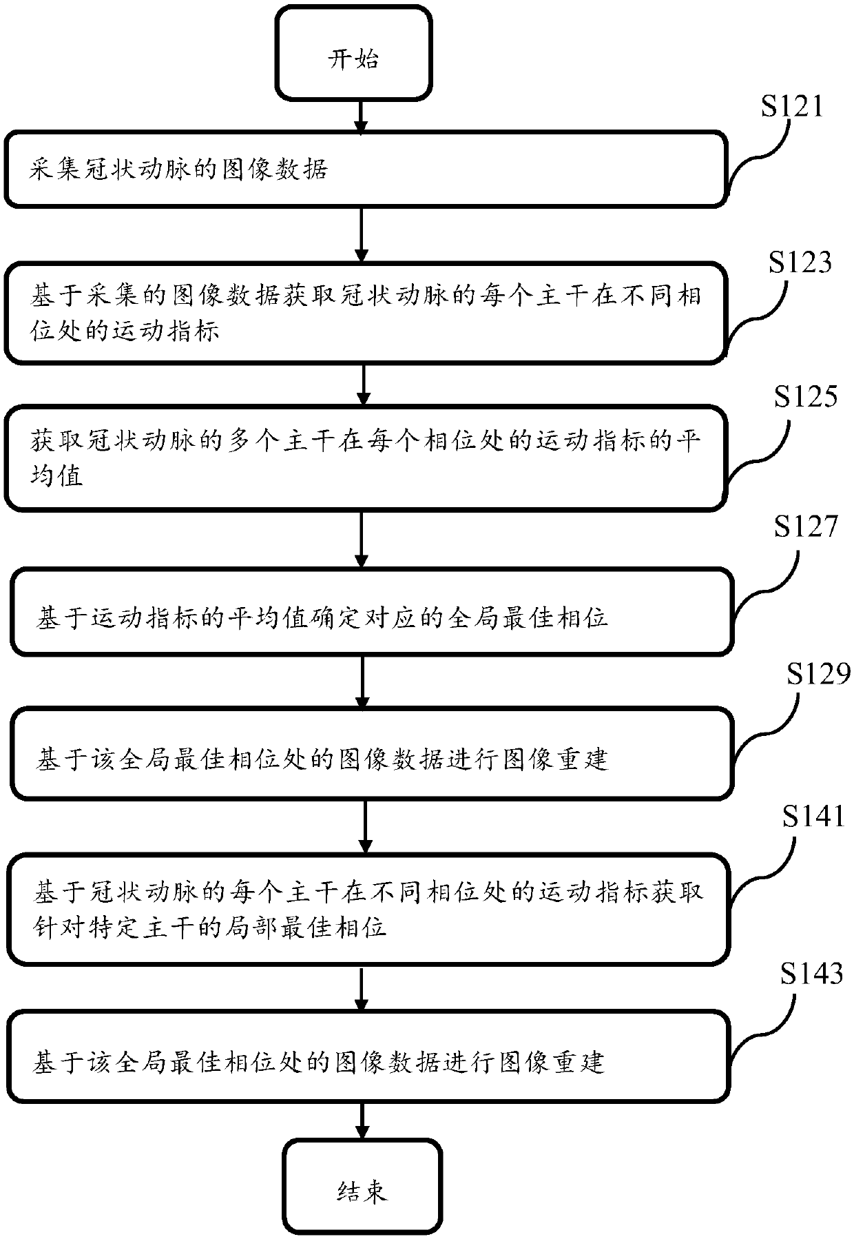
[0070] Among them, step 1 may include:
[0071] Sub-step 1-1: Collect image data of coronary arteries;
[0072] Sub-step 1-3: Obtain the motion index of each main stem of the coronary artery at different phases based on the collected image data;
[0073] Sub-step 1-5: Obtain the average value of the motion index of the multiple main trunks of t...
PUM
 Abstract
Description
Claims
Application Information
 - R&D
- Intellectual Property
- Life Sciences
- Materials
- Tech Scout
- Unparalleled Data Quality
- Higher Quality Content
- 60% Fewer Hallucinations
Browse by: Latest US Patents, China's latest patents, Technical Efficacy Thesaurus, Application Domain, Technology Topic, Popular Technical Reports.
© 2025 PatSnap. All rights reserved.Legal|Privacy policy|Modern Slavery Act Transparency Statement|Sitemap|About US| Contact US: help@patsnap.com


