
Urethrocystoscope system, urethrocystoscope sheath and obturator

A technology of cystoscope and obturator, which is applied in the field of urethral cystoscope system, urethral cystoscope sheath and obturator, which can solve the problems of inability to fill the urethra, loss of normal saline, difficulties in patient examination and diagnosis, and achieve observation Effects of eliminating blind spots, meeting diagnostic needs, and expanding uses

Pending Publication Date: 2021-04-02
泰州市高港中医院
View PDF0 Cites 0 Cited by
  • Summary
  • Abstract
  • Description
  • Claims
  • Application Information

AI Technical Summary

Problems solved by technology

In clinical work, the existing device can successfully complete most of the urethra and bladder inner wall observation, biopsy and other operations, but it cannot be used to observe and treat lesions within 3cm proximal to the male urethral orifice and female urethral distal end
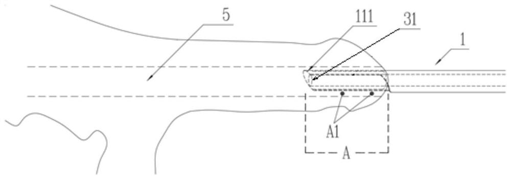
[0003] Tracing it to its cause is because the mirror sheath far-end 111 of existing urethrocystoscope system and the observation mirror 31 of endoscope far-end are on the same plane ( figure 2 ), and when observing 5 distal urethral lesions, the cystoscope sheath must be inserted into the patient’s urethra for more than 3cm to meet the requirements of the operation. The surgeon’s left hand must hold the head of the penis and insert a certain length of urethral cystoscope into the urethra At this time, the flushing water can enter the urethra and the situation in the distal urethra of the endoscope can be seen. Otherwise, the surgeon will not be able to hold the urethral cystoscope sheath with his left hand, and the cystoscope sheath may even fall off from the urethra. Examinations and operations that make it impossible to identify urethral diseases, but when the urethrocystoscope sheath is inserted into the urethra to a depth of more than 3 cm, see figure 2 The area A (urethra) within 3 cm from the proximal side of the external urethral opening is behind the observation mirror 31 on the distal end of the endoscope and becomes an observation blind spot, so that the existing urethrocystoscopy system cannot be used to observe and treat the male urethral opening Lesion A1 within 3cm proximally ( figure 2 )
In addition, when the scope sheath is inserted within 2.8cm, the perfused saline will also be lost from the semi-elliptical gap under the scope sheath, which cannot fill the urethra for observation
[0004] At present, the incidence of urethral lesions, especially lesions within 3 cm close to the external urethral orifice, has increased significantly. The above-mentioned status quo of urethral cystoscopy has brought difficulties to the examination and diagnosis of such patients. Therefore, it is necessary to design a method that can be used for A new type of urethrocystoscope sheath for observing and diagnosing lesions in this section of urethra

Method used


Image

  • Urethrocystoscope system, urethrocystoscope sheath and obturator
  • Urethrocystoscope system, urethrocystoscope sheath and obturator
  • Urethrocystoscope system, urethrocystoscope sheath and obturator

Examples


Embodiment 1

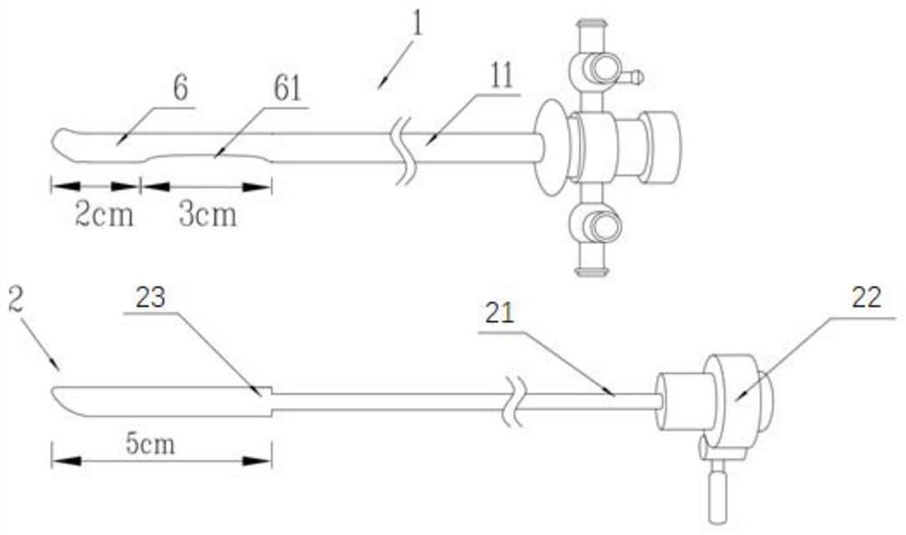
[0023] refer to image 3 with Figure 4 , a urethral cystoscope system, comprising: a mirror sheath 1, an obturator 2 that can be inserted into or pulled out of the mirror sheath 1, and an endoscope 3, and a conversion that can be connected to the mirror sheath 1 and the endoscope 3 respectively Connector 4, direction adjuster 8 and biopsy forceps 7, refer to image 3 and Figure 4 , the mirror sheath 1 includes a sheath tube 11, and the sheath tube 11 is an oblate tube. Different from the existing sheath tube, the sheath tube 11 of the present invention cancels the semicircle of 2.8 cm directly below the far end of the existing sheath tube. The gap is provided with a water injection valve and a water outlet valve at the proximal end of the sheath tube 11, and an extension connecting tube 6 is connected to the far end of the sheath tube 11 so that the operator can hold it without affecting operations such as observation and biopsy. An endoscope observation window 61 is prov...

Embodiment 2

[0025] A kind of urethrocystoscope sheath that described urethrocystoscope system uses, with reference to image 3 , including a sheath tube 11, a water injection valve and a water outlet valve are provided at the proximal end of the sheath tube 11, an extension connecting pipe 6 is connected at the far end of the sheath pipe 11, and an endoscope observation window 61 is provided on the extension connecting pipe 6 (refer to image 3 , Figure 4 or Figure 5 ), after the endoscope 3 is inserted into the sheath tube 11, the observation mirror 31 of the endoscope reaches the proximal edge of the observation window 61 of the endoscope. In this embodiment, the inner hole of the extension pipe 6 is a through hole, so that the distal end of the extension pipe 6 is open, the extension pipe 6 is flat and tubular, and the end nozzle of the extension pipe 6 is smooth and flat like a fish mouth. Garden shape; described endoscope observation window 61 begins at the 2cm place of described...

Embodiment 3

[0027] An obturator dedicated to the urethrocystoscope sheath of the urethrocystoscope system, refer to image 3 , comprising: a connecting rod 21, one end of the connecting rod 21 is provided with a sheath connector 22, and the other end of the connecting rod 21 is provided with a plunger 23, the shape of the plunger 23 is consistent with the extension of the urethrocystoscope sheath The inner shape of the tube is consistent, and the length of the plunger 23 is 5cm. The obturator is matched with the scope sheath and is used to close the end of the scope sheath tube and the 3cm gap on the tube wall, so as to avoid damage to the urethra during insertion.


PUM


Abstract

The invention discloses a urethrocystoscope system, a urethrocystoscope sheath and an obturator. The urethrocystoscope system comprises the sheath, the obturator capable of being inserted into or pulled out of the sheath, an endoscope, an adapter, a direction adjuster and biopsy forceps, wherein the adapter, the direction adjuster and the biopsy forceps can be connected with the sheath and the endoscope; the sheath comprises a sheath tube; a water injection valve and a water outlet valve are arranged at the near end of the sheath tube; an extension connecting tube is connected to the tail endof the sheath tube; a strip-shaped endoscope observation window is axially arranged at the near end of the extension connecting tube, namely the tail end of the sheath; an observation mirror of the endoscope inserted into the sheath abuts against the near side edge of the endoscope observation window; the sheath has the same shape as a sheath in an original urethrocystoscope system and is oblate;the obturator comprises a connecting rod; a sheath connector is arranged at one end of the connecting rod; a plunger is arranged at the other end of the connecting rod; and the shape of the plunger ismatched with the shape of the interior of the extension connecting tube of the urethrocystoscope sheath. According to the invention, the requirement that a urethrocystoscope is inserted into the urethra by 3 cm or above in an operation can be met, and full views of the urethra and the bladder within 3 cm away from the near side of the external urethral orifice can be observed.

Description

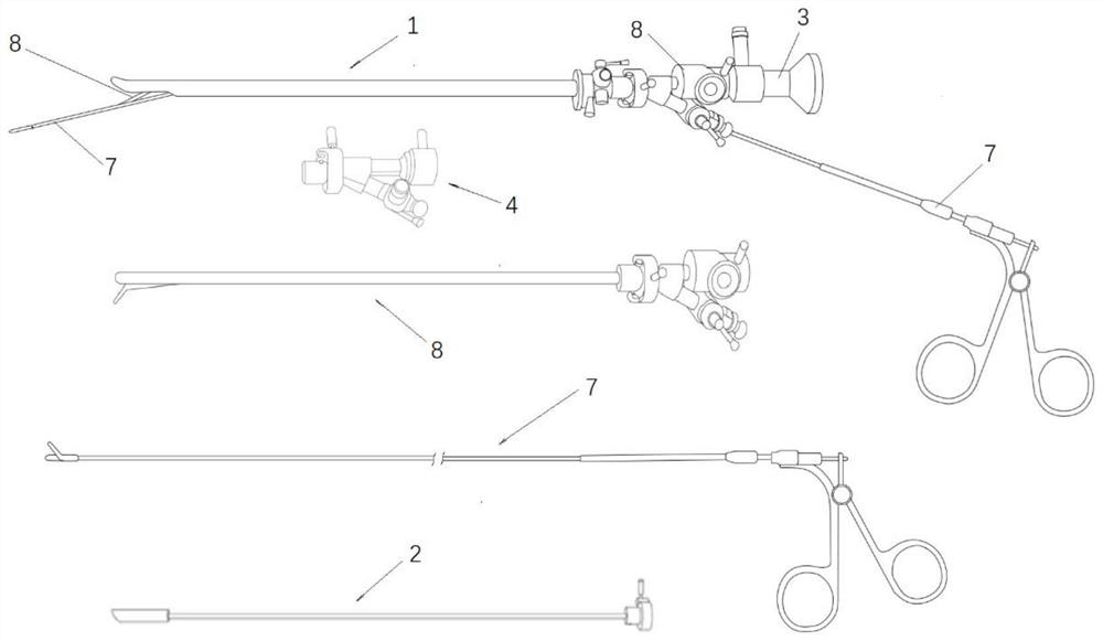
technical field [0001] The invention relates to a medical device, in particular to a urethrocystoscope system, a urethrocystoscope sheath and an obturator. Background technique [0002] The current clinically used urethral cystoscope system (refer to Fig. 1 ) is mainly composed of a scope sheath 1, an obturator 2, an endoscope 3 (respectively 0 degree, 30 degree, 70 degree and 120 degrees) and an adapter 4, Direction adjuster 8, biopsy forceps 7 etc. are formed. The scope sheath is 23cm long and has a flat garden shape. There is a semi-elliptical gap of about 2.8cm directly below the far side, which is convenient for inserting biopsy forceps or ureteral catheters during endoscopic observation. When in use, first insert the obturator into the urethrocystoscope sheath, apply lubricant to the outside of the sheath, pull out the obturator when inserted into the urethra for more than 3cm, and replace the assembled endoscope set with light source and flushing pipe , and then obs...

Claims


Application Information

Patent Timeline
Patent Type & Authority Applications(China)
IPC IPC(8): A61B1/307A61B1/015
CPCA61B1/015A61B1/307
Inventor 石广东孙建平吕春红许定珠
Owner 泰州市高港中医院
Learn More