Medical image detection method and device, equipment and storage medium

A medical image and detection method technology, applied in the field of image processing, can solve the problems of low robustness and low accuracy, and achieve the effect of high robustness and improved accuracy

Active Publication Date: 2019-07-09
SHANGHAI UNITED IMAGING INTELLIGENT MEDICAL TECH CO LTD
View PDF7 Cites 35 Cited by
  • Summary
  • Abstract
  • Description
  • Claims
  • Application Information

AI Technical Summary

Problems solved by technology

[0005] Based on this, it is necessary to address the technical problems of the low accuracy of the traditional medical image segmentation results and the low robustness of the traditional medical image segmentation algorithm. The present application provides a medical image detection method and device , equipment and storage media

Method used


Image

  • Medical image detection method and device, equipment and storage medium
  • Medical image detection method and device, equipment and storage medium
  • Medical image detection method and device, equipment and storage medium

Examples


Embodiment Construction

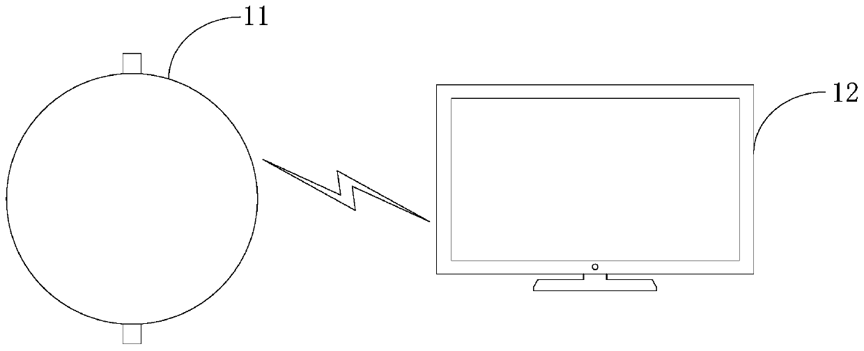
[0048] The medical image detection method provided in the present application is applicable to various medical image detection systems, such as MR systems, CT systems, PET systems, PET-CT systems, PET-MR systems, and ultrasound systems. figure 1 A schematic structural diagram of a medical image detection system provided for an embodiment, such as figure 1As shown, the system may include a scanning device 11 and a computer device 12, and the scanning device 11 and the computer device 12 may communicate in a wired manner or wirelessly. Optionally, the type of the scanning device 11 matches the type of the system to which it belongs, that is, when the system is an MR system, the scanning device 11 is an MR scanning device, and when the system is a PET system, the scanning device 11 is a PET detector, This embodiment does not limit the specific type of the scanning device 11 . Optionally, the computer device 12 may be an electronic device with a data processing function, such as ...


PUM


Abstract

The invention provides a medical image detection method and device, equipment and a storage medium. The method comprises the steps of obtaining a medical image; inputting the medical image into a segmentation model to obtain a target segmented image; wherein the target segmented image comprises a region of interest; inputting the target segmentation image and the medical image into a detection model to obtain a lesion detection image; wherein the lesion detection image comprises candidate positions of a lesion area, and the lesion area is located in the region of interest; wherein the segmentation model and the detection model are both deep learning models. The robustness of the segmentation model and the detection model provided by the method is high, the accuracy of the segmentation result of the medical image is improved, and the accuracy of the lesion detection result is improved.

Description

technical field [0001] The present application relates to the field of image processing, in particular to a medical image detection method, device, equipment and storage medium. Background technique [0002] At present, prostate cancer is the most common type of cancer that threatens the lives of millions of men around the world, and is the second leading cause of death among men. Therefore, early diagnosis and treatment of prostate cancer can greatly improve the survival rate and cure rate of patients. In the early diagnosis of prostate cancer, determining the location of the prostate from the patient's medical images is a crucial step. [0003] In traditional technology, a machine learning algorithm is usually used to segment medical images to obtain the location of the prostate. The specific process may be as follows: performing feature extraction on medical images, and then using a classifier to perform voxel-level classification on the extracted features, so as to dete...

Claims


Application Information

Patent Timeline
Patent Type & Authority Applications(China)
IPC IPC(8): G06T7/00G06T7/10G06T7/11
CPCG06T7/0012G06T2207/10081G06T2207/10088G06T2207/10104G06T2207/10132G06T2207/20081G06T2207/30096G06T7/10G06T7/11
Inventor 徐小栋廖术周翔詹翊强
Owner SHANGHAI UNITED IMAGING INTELLIGENT MEDICAL TECH CO LTD
Why Eureka
Try Eureka