Femoral head CT image segmentation method

A CT image and femoral head technology, applied in the field of medical image processing, can solve problems such as low segmentation accuracy, over-segmentation, and impact on segmentation results

Active Publication Date: 2019-10-18
NORTHEASTERN UNIV
View PDF4 Cites 5 Cited by
  • Summary
  • Abstract
  • Description
  • Claims
  • Application Information

AI Technical Summary

Problems solved by technology

Among them, the threshold method relies solely on image pixel information for segmentation, which has the disadvantages of ignoring image noise and low contrast at the boundary. When segmenting CT images of the femoral head, the outline of the femoral head will be incomplete, there will be large cavities inside, and there will be too many bright spots of noise. Problem; the watershed method will be affected by the detailed texture and noise in the CT image, and over-segmentation will occur, which will affect the segmentation effect and have a high mis-segmentation rate; the level set method has strict requirements for the selection of the initial point, and the segmentation speed is slow and the segmentation is accurate The rate is low; the atlas method requires a large number of training samples, and when the difference between the training samples and the test samples is too large, accurate segmentation cannot be completed

Method used


Image

  • Femoral head CT image segmentation method
  • Femoral head CT image segmentation method
  • Femoral head CT image segmentation method

Examples


Embodiment Construction

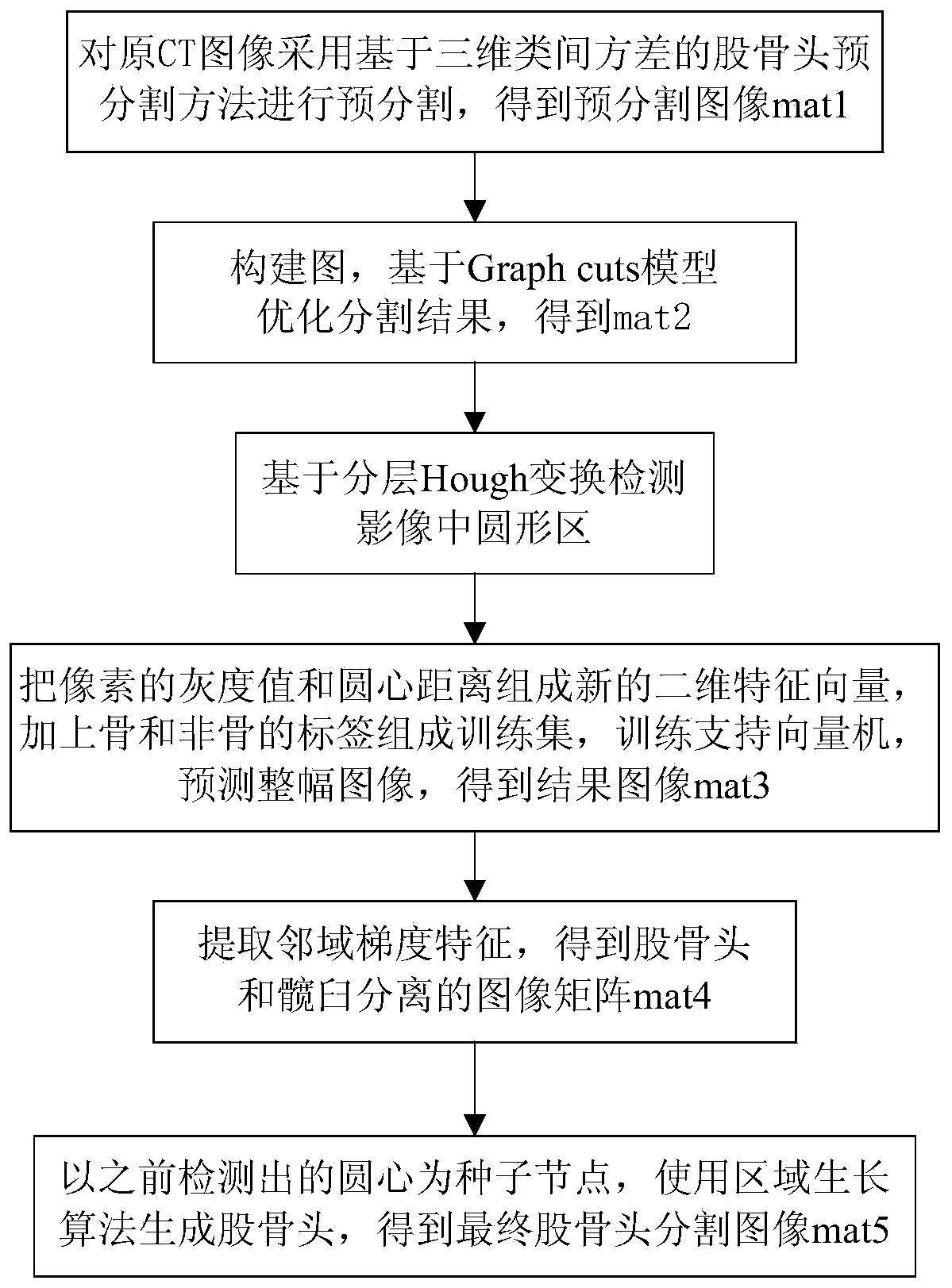
[0047] The specific implementation manners of the present invention will be further described in detail below in conjunction with the accompanying drawings and embodiments. The following examples are used to illustrate the present invention, but are not intended to limit the scope of the present invention.

[0048] Such as figure 1 As shown, the method of this embodiment is as follows.

[0049] Step 1: Femoral head pre-segmentation method based on 3D inter-class variance;

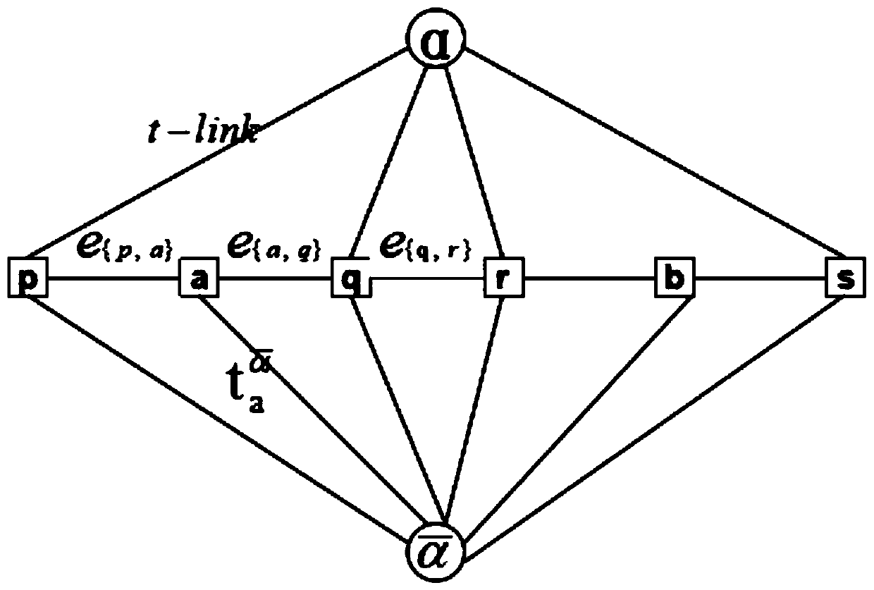
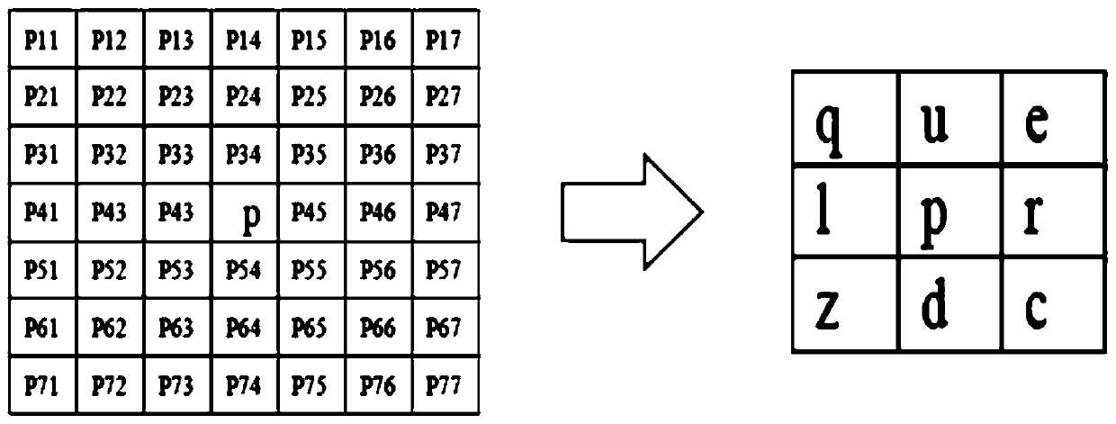
[0050] Since the segmentation method based on the Graph Cuts model is an interactive segmentation method, the user needs to mark some pixels as "object" or "background" in advance to provide hard constraints for segmentation, so the three-dimensional maximum between-class variance method is used for Pre-segmentation. According to the segmentation results, the 10% pixels with the highest gray value in the bone pixel set and the 10% non-zero pixels with the lowest gray value in the non-bone pixel set are ta...


PUM


Abstract

The invention provides a femoral head CT image segmentation method, and relates to the technical field of medical image processing. The method comprises: performing pre-segmentation by using a three-dimensional maximum between-cluster variance method; then, based on the combination of image segmentation and shape constraint, performing automatic femoral head accurate segmentation; after graph construction, optimizing a segmentation result on the basis of a Graph cuts model; detecting a circular area in the image based on layered Hough transform; and carrying out re-prediction and classification on a Graph cuts segmentation result by using an SVM, extracting neighborhood gradient features, separating the femoral head from the acetabulum, and generating the femoral head by using a regional growth algorithm by taking the detected circle center as a seed node to obtain a final femoral head segmentation image. According to the invention, image noise can be effectively eliminated, hard constraint conditions are provided for the Graph cuts model, robustness is good, full-automatic segmentation of the femoral head CT image is achieved, the convergence time of the Graph cuts model can be greatly shortened, the edges of the segmented femoral head are complete, details are clear, and the segmentation accuracy reaches 92%.

Description

technical field [0001] The invention relates to the technical field of medical image processing, in particular to a method for segmenting a CT image of a femoral head. Background technique [0002] Image segmentation plays a very important role in the quantitative and qualitative analysis of medical images, and it directly affects the subsequent analysis and processing of computer-aided diagnosis systems. Correctly segmenting the image of the femoral head can not only determine the degree of necrosis of the patient through the shape of the femoral head, but also approximate the ischemic volume inside the patient's femoral head through the segmentation results, which can be used for auxiliary diagnosis and stage judgment of femoral head necrosis. get ready. At present, the segmentation methods of femoral head CT images mainly include expert manual segmentation, computer interactive segmentation and automatic segmentation. Manual segmentation and computer interactive segment...

Claims


Application Information

Patent Timeline
Patent Type & Authority Applications(China)
IPC IPC(8): G06T7/11G06K9/62
CPCG06T7/11G06T2207/10081G06T2207/30008G06F18/2411G06F18/24323
Inventor 栗伟于鲲冯朝路王东杰覃文军赵大哲
Owner NORTHEASTERN UNIV
Why Eureka
Try Eureka