Biological active scaffold and preparation method thereof
A bioactive and buffer technology, applied in the field of micro/nano bioactive scaffolds with supramolecular recognition functions, can solve the problems of difficulty in ensuring activity and lack of biological carriers, and achieve the effects of promoting repair, preventing cerebrospinal fluid leakage, and promoting migration behavior
- Summary
- Abstract
- Description
- Claims
- Application Information
AI Technical Summary
Problems solved by technology
Method used
Image

Examples
Embodiment 1
[0036] A bioactive scaffold includes the following components: silk fibroin and Col-I, and the mass ratio of the silk fibroin to Col-I is 15:1.
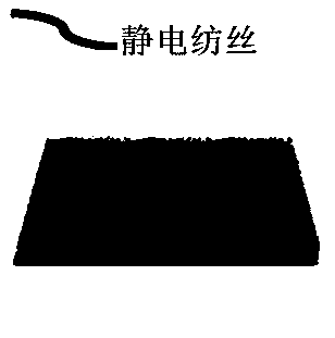
[0037] The dry silk fibroin was dissolved in 7% w / t hexafluoroisopropanol to obtain a silk fibroin solution, and the silk fibroin solution was electrospun at an injection speed of 0.8 ml / min, a voltage of 12 kV, and a jet collection distance of 10 cm Silk treatment, after electrospinning, soak in anhydrous methanol, wash with deionized water, and freeze-dry to obtain SF electrospun membrane. Add the SF electrospun membrane to the tris-HCl buffer solution of dopamine (concentration 2 mg / ml, pH=8.5) and soak for 12 h.
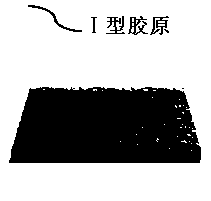
[0038] Dissolve Col-I in 0.006 mmol / L acetic acid at a concentration of 3 mg / L, mix it with 10×PBS solution at a ratio of 1:6, and adjust the pH to 7.0.
[0039] Drop Col-I onto the SF electrospun membrane, place it at 37°C for 30 min, and wash it with deionized water to obtain the Col-I modified SF electrospun membran...
Embodiment 2
[0042] A bioactive scaffold includes the following components: silk fibroin and Col-I, and the mass ratio of the silk fibroin to Col-I is 15:1.
[0043]The silk fibroin solution was obtained by dissolving the dry silk in 6% w / t hexafluoroisopropanol, and the silk fibroin solution was subjected to electrospinning treatment at an injection speed of 0.6ml / min, a voltage of 15 kV, and a spray collection distance of 15 cm. After spinning, soak in anhydrous methanol, wash with deionized water, and freeze-dry to obtain SF electrospun membrane. The SF electrospun membrane was soaked in tris-HCl buffer solution of dopamine (concentration 3 mg / ml, pH=8.6) for 12 h.
[0044] Dissolve Col-I in 0.009 mmol / L acetic acid at a concentration of 5 mg / L, mix it with 10×PBS solution at a ratio of 1:6, and adjust the pH to 7.0.
[0045] Drop Col-I onto the SF electrospun membrane, place it at 37°C for 30 min, and wash it with deionized water to obtain the Col-I modified SF electrospun membrane, w...
Embodiment 3
[0047] A bioactive scaffold includes the following components: silk fibroin and Col-I, and the mass ratio of the silk fibroin to Col-I is 15:1.
[0048] The silk fibroin solution was obtained by dissolving the dry silk in 9% w / t hexafluoroisopropanol. The silk fibroin solution was subjected to electrospinning at a sampling rate of 1.0ml / min, a voltage of 16 kV, and a jet collection distance of 8 cm. After spinning, soak in anhydrous methanol, wash with deionized water, and freeze-dry to obtain SF electrospun membrane. The SF electrospun membrane was soaked in tris-HCl buffer solution of dopamine (concentration 1 mg / ml, pH=8.2) for 12 h.
[0049] Dissolve Col-I in 0.003mmol / L acetic acid at a concentration of 1 mg / L, mix it with 10×PBS solution at a ratio of 1:6, and adjust the pH to 7.0.
[0050] Drop Col-I onto the SF electrospun membrane, place it at 37°C for 30 min, and wash it with deionized water to obtain the Col-I modified SF electrospun membrane, wherein the mass rati...
PUM
| Property | Measurement | Unit |
|---|---|---|
| Concentration | aaaaa | aaaaa |
Abstract
Description
Claims
Application Information
 - R&D Engineer
- R&D Manager
- IP Professional
- Industry Leading Data Capabilities
- Powerful AI technology
- Patent DNA Extraction
Browse by: Latest US Patents, China's latest patents, Technical Efficacy Thesaurus, Application Domain, Technology Topic.
© 2024 PatSnap. All rights reserved.Legal|Privacy policy|Modern Slavery Act Transparency Statement|Sitemap