
Biological sample tensile test method

A technique for stretch testing and biological samples, applied in the field of biotechnology research, can solve the problem of cell dynamic stretch limitation, not suitable for live cell in situ imaging, adjustable frequency range, adjustable stretch amplitude range cannot meet the experimental requirements And other problems, to achieve the effect of not easy to tear, large amplitude range, large adjustable range

Active Publication Date: 2019-10-25
JINHUA VOCATIONAL TECH COLLEGE
View PDF14 Cites 0 Cited by
  • Summary
  • Abstract
  • Description
  • Claims
  • Application Information

AI Technical Summary

Problems solved by technology

[0002] In the process of cell tissue development and repair, mechanical stretching can be used as an important means to regulate the state and function of cells. There are many methods in the prior art to mechanically stimulate cells, but most of them use mechanical actuation. The existing technology of stretching device is not suitable for in situ imaging of live cells at the subcellular scale, and among the few existing technologies capable of real-time live cell imaging at the subcellular scale, the dynamic stretching of cells is limited , and its adjustable frequency range and adjustable stretching amplitude range cannot meet the experimental requirements, the biological sample stretching test method can solve the problem

Method used


Image

  • Biological sample tensile test method
  • Biological sample tensile test method

Examples


Embodiment Construction

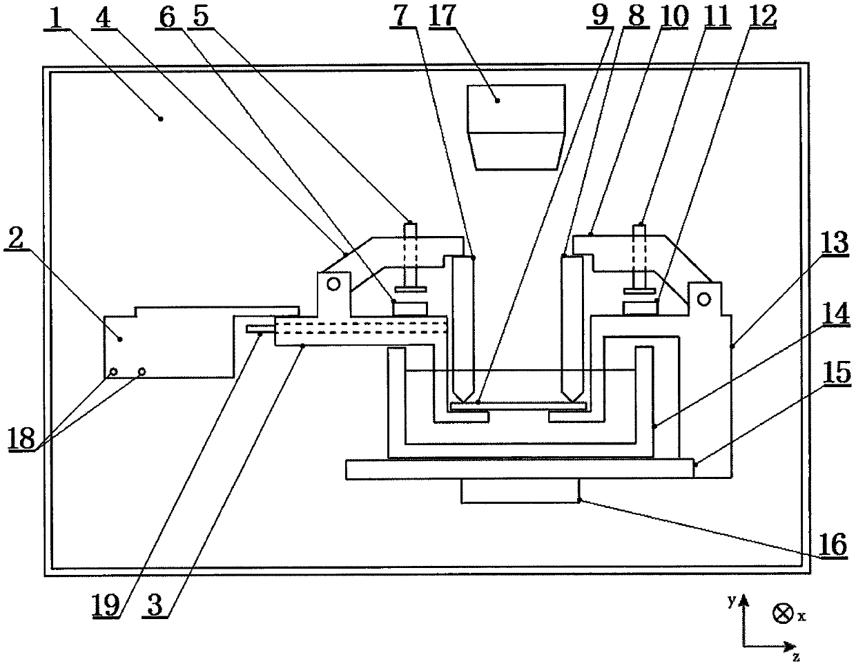
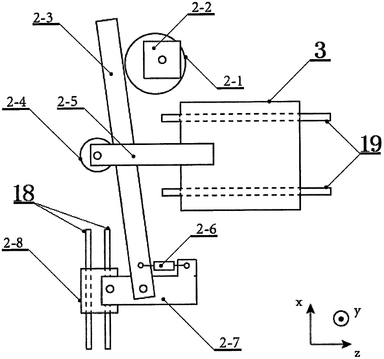
[0021] Such as figure 1 It is a schematic diagram of the present invention, including a housing 1 and a driver 2 located in the housing 1, a movable clamp 3, a movable arm I4, an adjustment rod I5, a magnet I6, a vertical clamp I7, a vertical clamp II8, a substrate 9, a movable arm II10, Adjusting rod II11, magnet II12, fixing fixture 13, liquid reservoir 14, support platform 15, displacement stage 16, microscope 17, guide rail I18 and guide rail II19, xyz is a three-dimensional coordinate system, and shell 1 is a metal cavity in the shape of a cuboid. 1 has several operating windows, the protective liquid suitable for the cell culture environment is housed in the liquid reservoir 14, the microscope 17 is connected with the computer through the cable, and the displacement platform 16, two parallel guide rails I18 and two parallel guide rails II19 are all fixed on the In the shell 1, the guide rail I18 and the guide rail II19 are vertical in space, the support platform 15 is lo...


PUM

PropertyMeasurementUnit
Diameteraaaaaaaaaa

Abstract

The invention relates to the field of biotechnology research, and discloses a biological sample tensile test method. The biological sample tensile test method adopts a biological sample tensile test device, which comprises an outer shell, and a driver, a movable clamp, a movable arm I, an adjusting rod I, a magnet I, a vertical clamp I, a vertical fixture II, a substrate, a movable arm II, an adjusting rod II, a magnet II, a fixed fixture, a liquid storage tank, a supporting stage, a displacement stage, a microscope, a guide rail I and a guide rail II which are arranged inside the outer shell.The biological sample tensile test method can carry out in-situ imaging on subcellular-scale live cells, can perform dynamic tension at the same time, adopts the special driving mechanism and clamp structures, can perform single-axis tension on a biological cell sample while conducting in-situ imaging on the subcellular-scale live cells, so as to study properties of the cells after tension stimulus through in-situ imaging, is not prone to cause fracture of the substrate, and has wide adjustable range of tension frequency and amplitude and wide adjustable amplitude range of tension.

Description

technical field [0001] The invention relates to the field of biotechnology research, in particular to a biological sample tensile test method capable of in situ imaging of subcellular scale live cells and capable of dynamic stretching at the same time. Background technique [0002] In the process of tissue development and repair of cells, mechanical stretching can be used as an important means to regulate the state and function of cells. There are many methods in the prior art to mechanically stimulate cells, but most of them use mechanical actuation. The existing technology of stretching device is not suitable for in situ imaging of live cells at the subcellular scale, and among the few existing technologies capable of real-time live cell imaging at the subcellular scale, the dynamic stretching of cells is limited , and its adjustable frequency range and adjustable stretching amplitude range cannot meet the experimental requirements, the biological sample stretching test me...

Claims


Application Information

Patent Timeline
IPC IPC(8): G01N19/00G01N21/84
CPCG01N19/00G01N21/84
Inventor 索奕双郭强张向平
Owner JINHUA VOCATIONAL TECH COLLEGE
Learn More