
Visual device for intraoperative anastomotic bleeding diagnosis

An anastomosis and rod placement technology, applied in the directions of diagnosis, diagnostic recording/measurement, application, etc., can solve problems such as life-threatening, poor suture quality, anastomotic bleeding, etc., and achieve the effect of easy viewing

Pending Publication Date: 2019-11-15
JIANGSU CANCER HOSPITAL
View PDF0 Cites 0 Cited by
  • Summary
  • Abstract
  • Description
  • Claims
  • Application Information

AI Technical Summary

Problems solved by technology

[0002] At present, after lumen anastomosis operations of the digestive tract and urinary tract such as esophagus, gastrointestinal tract, pancreas, biliary tract, and ureter, it is usually impossible to effectively expose and observe the reconstructed anastomotic stoma from the lumen due to various reasons. It is very convenient and quick to carry out special quality inspection tools for the anastomotic stoma after suturing, and only rely on the doctor's clinical experience to control it. For some young doctors with less work experience, there are serious problems such as anastomotic bleeding and anastomotic leakage after surgery. risk of complications
At the same time, even doctors with rich clinical experience will inevitably be negligent. Poor suture quality will often lead to local bleeding, hematoma or leakage of the anastomotic stoma, resulting in postoperative anastomotic bleeding, slow healing, or even failure to heal, resulting in anastomotic leakage, etc. Severe and fatal complications often require re-examination, interventional treatment and even re-operation at great risk, which brings great pain to the patient and even endangers his life

Method used


Image

  • Visual device for intraoperative anastomotic bleeding diagnosis
  • Visual device for intraoperative anastomotic bleeding diagnosis
  • Visual device for intraoperative anastomotic bleeding diagnosis

Examples


Embodiment 1

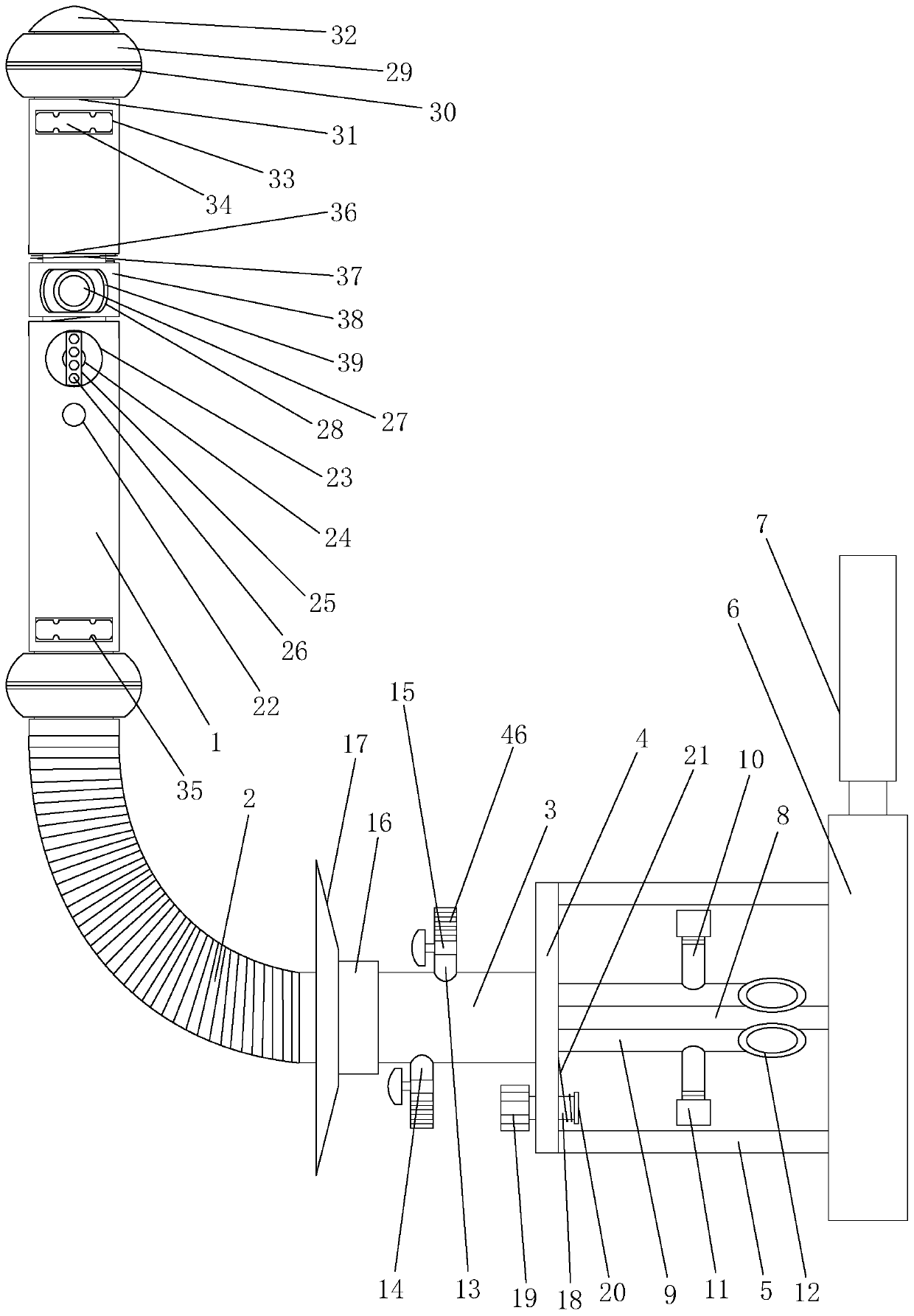
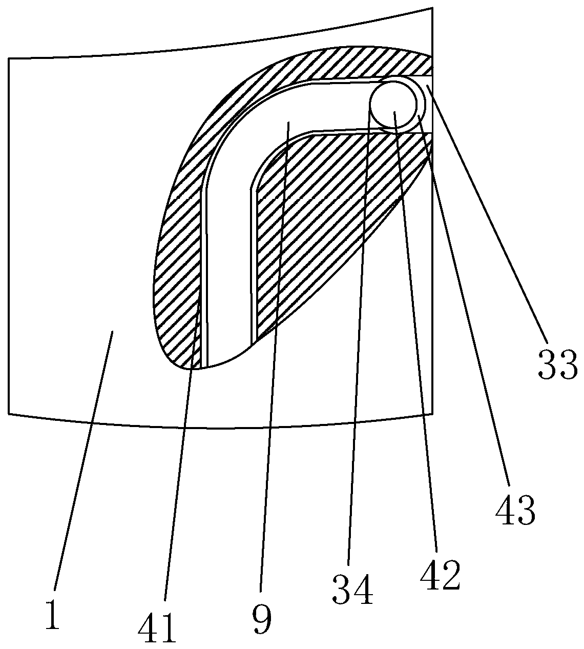
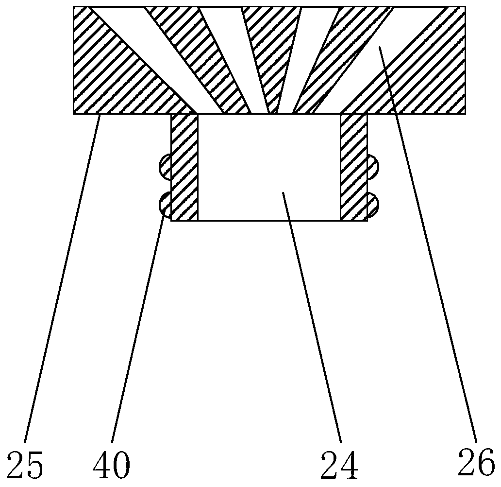
[0023] Such as Figure 1-5 As shown, the visual device for diagnosis of anastomotic bleeding in the operation disclosed by the present invention includes: an insertion rod 1, a bent bellows 2, an external rod 3, a terminal bracket and a display terminal 6; An annular airbag 29 is installed at each end; a camera 27 and a lighting lamp 22 are installed on the wall of the middle part of the insertion rod 1; One end of the terminal is butt-connected and installed; the display terminal 6 is installed on the other end of the external pole 3 through the terminal bracket, and the display terminal 6 is electrically connected with the camera 27 for real-time display of the captured images of the camera 27; Extending to the wire hole at the camera 27 and the lighting lamp 22, a wire tube through hole is provided on the external rod 3 along its axial direction; an insulating wire tube 8 is installed on the wire hole, and the other end of the insulating wire tube 8 is sequentially penetrat...


PUM


Abstract

The invention provides a visual device for intraoperative anastomotic bleeding diagnosis. The device comprises an insertion rod, a bent corrugated pipe, an external rod, a terminal bracket and a display terminal; the head and tail ends of the insertion rod are provided with annular airbags respectively; the middle rod wall of the insertion rod is provided with a camera and an illumination lamp; the two ends of the bent corrugated pipe are in butt joint with one end of the insertion rod and one end of the external rod respectively. The visual device for the intraoperative anastomotic bleeding diagnosis can collect clear images to the display terminal for display by using the camera and the illumination lamp, thereby providing convenience for a doctor to check whether or not bleeding occursat an anastomotic stoma; the two ends of an insertion cavity can be conveniently closed by using the annular airbags arranged at the two ends, and the cavity can also be propped up, thereby providingconvenience for checking the anastomotic stoma.

Description

technical field [0001] The invention relates to a visual device, in particular to a visual device for diagnosing anastomotic bleeding in operation. Background technique [0002] At present, after lumen anastomosis operations of the digestive tract and urinary tract such as esophagus, gastrointestinal tract, pancreas, biliary tract, and ureter, it is usually impossible to effectively expose and observe the reconstructed anastomotic stoma from the lumen due to various reasons. It is very convenient and quick to carry out special quality inspection tools for the anastomotic stoma after suturing, and only rely on the doctor's clinical experience to control it. For some young doctors with less work experience, there are serious problems such as anastomotic bleeding and anastomotic leakage after surgery. risk of complications. At the same time, even doctors with rich clinical experience will inevitably be negligent. Poor suture quality will often lead to local bleeding, hematoma ...

Claims


Application Information

Patent Timeline
Patent Type & Authority Applications(China)
IPC IPC(8): A61B5/02
CPCA61B5/02042
Inventor 陈天鸣陈仕林潘耀振
Owner JIANGSU CANCER HOSPITAL
Learn More