Ultrasound Imaging Catheter With Pivoting Head

a technology of ultrasound imaging and pivoting head, which is applied in the field of catheters, can solve the problems of limited three-dimensional viewing perspective, and achieve the effect of greater viewing perspectiv

Active Publication Date: 2009-10-22
ST JUDE MEDICAL ATRIAL FIBRILLATION DIV
View PDF52 Cites 193 Cited by
  • Summary
  • Abstract
  • Description
  • Claims
  • Application Information

AI Technical Summary

Benefits of technology

[0007]The various embodiments provide an ultrasound imaging catheter with a pivoting head portion which enables the ultrasound imaging transducer array to be pivoted about a hinge with a near-zero radius of curvature. The pivot motion of the ultrasound imaging transducer array is controlled from a handle, such as by pivot cables connected to a control wheel in the handle. Pivoting the ultrasound imaging transducer array through a large angle, such as 90° from the centerline of the catheter, enables clinicians to obtain intracardiac ultrasound images from different perspectives without repositioning the catheter itself. In an embodiment, the ultrasound imaging transducer array is capable of being pivoted plus and minus 90° from a center line position (i.e., parallel to the main axis of the catheter). In an embodiment, the catheter may also be steerable including a capability of bending a distal portion of the catheter through a finite radius of curvature by steering cables controlled from the handle. By combining bending of the catheter with pivoting of the transducer head, greater viewing perspectives can be provided. Embodiments include a handle assembly featuring two control actuators for controlling the steering and pivoting mechanisms of the attached catheter. The various embodiments are not necessarily limited to ultrasound imaging catheters, and may be implemented in any catheter that may benefit from having a distal tip capable of pivoting about a hinge.

Problems solved by technology

As the transducer face is linear and parallel to the catheter shaft, and because the two directional controls are limited, the, current design steerable ultrasound catheters provide a limited three-dimensional viewing perspective.

Method used


Image

  • Ultrasound Imaging Catheter With Pivoting Head
  • Ultrasound Imaging Catheter With Pivoting Head
  • Ultrasound Imaging Catheter With Pivoting Head

Examples


Embodiment Construction

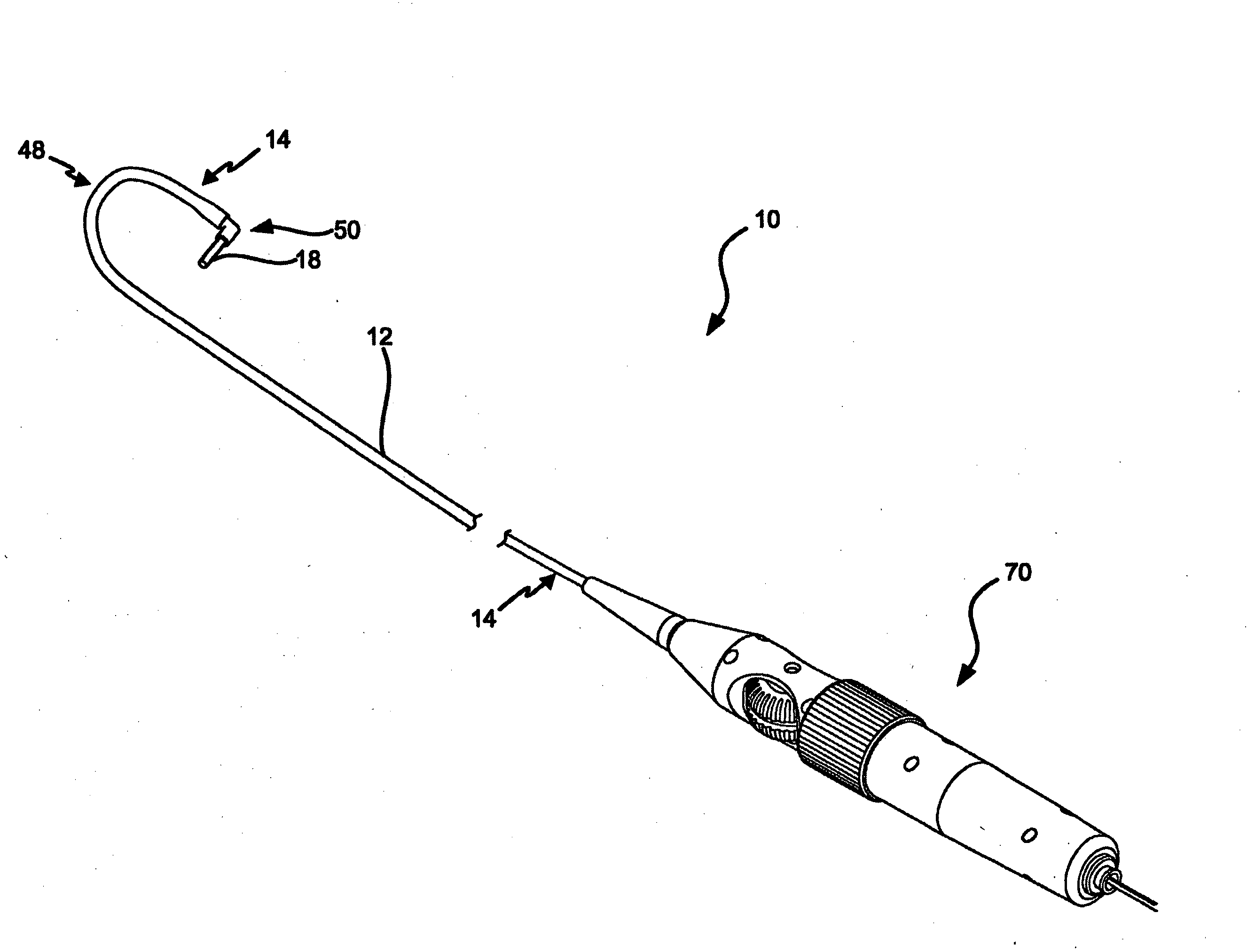
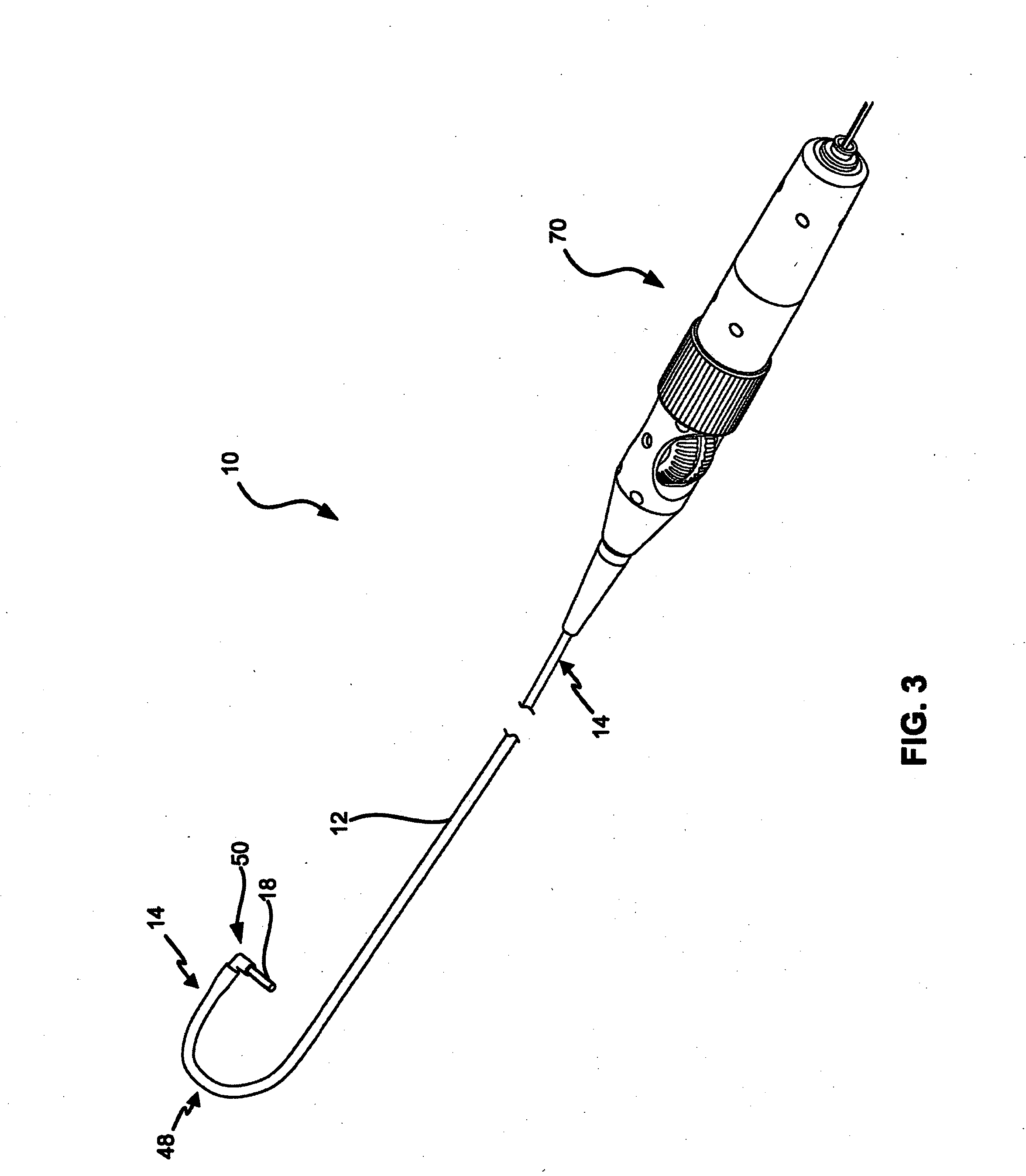
[0025]The various embodiments will be described in detail with reference to the accompanying drawings. Wherever possible, the same reference numbers will be used throughout the drawings to refer to the same or like parts. References made to particular examples and implementations are for illustrative purposes, and are not intended to limit the scope of the invention or the claims.

[0026]As used herein, the terms “about” or “approximately” for any numerical values or ranges indicates a suitable dimensional tolerance that allows the part or collection of components to function for its intended purpose as described herein. Also, as used herein, the terms “patient,”“host” and “subject” refer to any human or animal subject and are not intended to limit the systems or methods to human use, although use of the subject invention in a human patient represents a preferred embodiment.

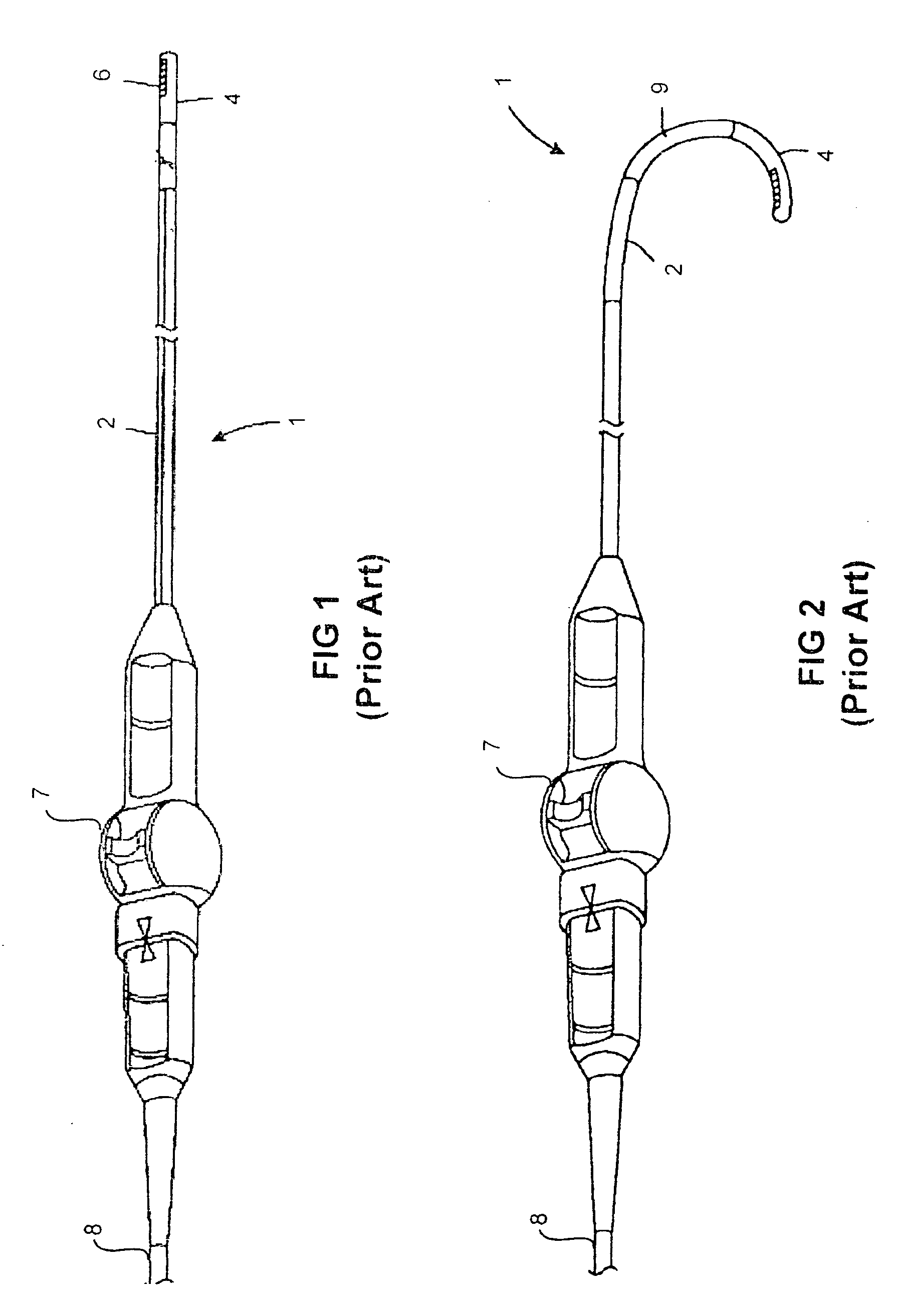
[0027]As used herein, the term “catheter” is used as a general reference to any elongated tubular assembly which...


PUM


Abstract

An ultrasound imaging catheter system includes a pivot head assembly coupled between a ultrasound transducer array and the distal end of the catheter. The pivot head assembly includes a pivot joint enabling the transducer array to pivot through a large angle about the catheter centerline in response pivot cables controlled by a wheel within a handle assembly. Pivoting the ultrasound transducer array approximately 90° once the catheter is positioned by rotating the catheter shaft and bending the distal section of the catheter, clinicians may obtain orthogonal 2-D ultrasound images of anatomical structures of interest in 3-D space. Combining bending of the catheter by steering controls with pivoting of the transducer head enables a greater range of viewing perspectives. The pivot head assembly enables the transducer array to pan through a large angle to image of a larger volume than possible with conventional ultrasound imaging catheter systems.

Description

FIELD OF THE INVENTION[0001]The present invention relates generally to catheters and more particularly to a steerable ultrasound imaging catheter with a pivoting head.BACKGROUND[0002]Catheters for insertion and deployment within blood vessels and cardiac chambers are well-known in the art. A variety of catheters are now utilized in the diagnosis and treatment of cardiac disease. Among the ensemble of catheters used in cardiac care are ultrasound imaging catheters which can be inserted within the heart (i.e., intracardial) to obtain two-dimensional ultrasound images of heart structures and measure blood flow. Such intracardial echocardiography (ICE) catheters provide cardiologist and heart surgeons with unique viewing perspectives beneficial to diagnosis and treatment of heart diseases.[0003]To obtain diagnostically useful images, an ultrasound imaging catheter can be positioned next to or within the vessels and chambers of the heart. Typically, the catheter is introduced into a pati...

Claims


Application Information

Patent Timeline
Patent Type & Authority Applications(United States)
IPC IPC(8): A61B8/14A61B8/12
CPCA61B8/0883A61B8/12A61M25/0136A61B8/4466A61B8/4488A61B8/445
Inventor BYRD, CHARLES BRYAN
Owner ST JUDE MEDICAL ATRIAL FIBRILLATION DIV
Why Eureka
Try Eureka