Fundus image blood vessel segmentation method based on Frangi enhancement and attention mechanism UNet

A fundus image and attention technology, applied in the field of image analysis and deep learning, can solve problems such as uneven illumination, blur, noise, etc., to achieve the effect of increasing continuity and integrity, strong generalization ability, and improving contrast.

Active Publication Date: 2019-11-19
FUZHOU UNIV
View PDF7 Cites 23 Cited by
  • Summary
  • Abstract
  • Description
  • Claims
  • Application Information

AI Technical Summary

Problems solved by technology

However, the current method still has the following limitations: (1) It is easily affected by non-vascular factors in the fundus image, such as noise, lesions and illumination, resulting in poor segmentation of blood vessels; (2) The generalization ability of the vascular segmentation model Poor, some models perform well on a single dataset, but split poorly when tested on multiple datasets or between different datasets
[0004] For the blood vessel segmentation of fundus images, there are mainly two problems: on the one hand, there are often various imaging artifacts caused by insufficient light during fundus imaging, such as blurring, noise, uneven illumination, etc. There is also the influence of pathological factors in fundus images, which makes it difficult to segment blood vessels, especially the segmentation of small blood vessels; on the other hand, most of the existing depth models only perform algorithm design on a single or two data sets. Limited generalization ability

Method used


Image

  • Fundus image blood vessel segmentation method based on Frangi enhancement and attention mechanism UNet
  • Fundus image blood vessel segmentation method based on Frangi enhancement and attention mechanism UNet
  • Fundus image blood vessel segmentation method based on Frangi enhancement and attention mechanism UNet

Examples


Embodiment Construction

[0047] The present invention will be further described below in conjunction with the accompanying drawings and embodiments.

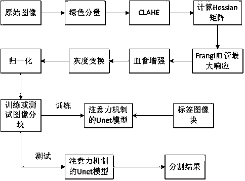
[0048] like figure 1 As shown, the present embodiment provides a fundus image blood vessel segmentation method based on Frangi enhancement and attention mechanism UNet, comprising the following steps:

[0049] Step S1: Provide an RGB fundus image as an input image, extract a green component from the input image, and use the contrast-limited histogram equalization method (CLAHE) to perform contrast adjustment on the image after the green component is extracted;

[0050] Step S2: Calculate the Hessian matrix of each pixel in the image after the contrast adjustment in step S1, and obtain the eigenvalue of the Hessian matrix;

[0051] Step S3: using the eigenvalues ​​of the Hessian matrix to construct a Frangi vessel similarity function under the condition that the scale factor is σ, and obtain the maximum response;

[0052] Step S4: Subtract the product ...


PUM


Abstract

The invention relates to a fundus image blood vessel segmentation method based on Frangi enhancement and an attention mechanism UNet, and the method comprises the steps: firstly extracting a green component from an input image, and carrying out the contrast adjustment on the basis of the extracted green component through a contrast-limited histogram equalization method; calculating a Hessian matrix of each pixel point in the image after the contrast ratio is adjusted; constructing a Frangi vascular similarity function by utilizing the characteristic value of the Hessian matrix under the condition of a scale factor, and obtaining the maximum response; respectively subtracting the product of the maximum response value and the enhancement factor factor factor from the pixel values of the RGBthree same channels of each pixel point of the input image; then, carrying out gray scale transformation on the image after frangi enhancement, and carrying out zero mean normalization operation on each pixel value to be between [0, 1]; and finally, inputting the obtained training image blocks and label image blocks into an attention mechanism UNet network for training; and obtaining a segmentation result through testing. According to the invention, the generalization ability of the model is improved.

Description

technical field [0001] The invention relates to the technical field of image analysis and deep learning, in particular to a fundus image blood vessel segmentation method based on Frangi enhancement and attention mechanism UNet. Background technique [0002] Fundus blood vessels are the only part of the human blood circulation system that can be directly observed non-invasively. Studies have shown that fundus vascular abnormalities are related to the existence and severity of coronary heart disease, hypertension, diabetes, atherosclerosis, and kidney disease. For example, the improvement rate of localized retinal artery stenosis is closely related to the degree of hypertension control. The extraction and measurement of blood vessels in fundus images has important clinical significance for the auxiliary detection and quantitative analysis of related diseases, and the accurate segmentation of blood vessels is the premise of the above work. Due to individual differences, there ...

Claims


Application Information

Patent Timeline
Patent Type & Authority Applications(China)
IPC IPC(8): G06T7/00G06T7/11G06T5/00G06N3/04G06N3/08
CPCG06T7/0012G06T7/11G06T5/007G06N3/08G06T2207/30041G06T2207/20081G06T2207/20084G06N3/045
Inventor 潘林朱有煌
Owner FUZHOU UNIV
Why Eureka
Try Eureka