Method and equipment for detecting tissue elasticity

An elastic detection and tissue technology, applied in the field of medical imaging, can solve the problems of detection error, inability to know the tissue structure information of the detection area, etc., and achieve the effect of improving the accuracy

Active Publication Date: 2019-12-27
WUXI HISKY MEDICAL TECH
View PDF1 Cites 1 Cited by
  • Summary
  • Abstract
  • Description
  • Claims
  • Application Information

AI Technical Summary

Problems solved by technology

[0004] However, the transient elastography technique cannot obtain the tissue structure information of the detection area, especially the two-dimensional structure information of the tissue, and the technician usually can only set and arrange the ultrasonic probe for transient elastography based on experience
Therefore, when performing tissue elasticity detection, the ultrasonic probe detects the detection area to generate images, but due to the movement of elastic tissue caused by breathing, heartbeat, etc., and when there may be large blood vessels, cysts, or ascites in the area to be inspected, etc. Detection errors cannot be avoided. When selecting a detection area, consider how to avoid the above situation and choose a detection area that is less affected
[0005] In the related technology, in the process of tissue elasticity detection, how to select the detection area with less influence of tissue dynamics, no effective solution has been proposed so far.

Method used


Image

  • Method and equipment for detecting tissue elasticity
  • Method and equipment for detecting tissue elasticity
  • Method and equipment for detecting tissue elasticity

Examples


Embodiment Construction

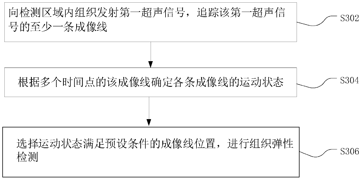
[0033] In order to make the purpose, technical solution and advantages of the present application clearer, the present application will be further described in detail below in conjunction with the accompanying drawings and embodiments. It should be understood that the specific embodiments described here are only used to explain the present application, and are not intended to limit the present application.

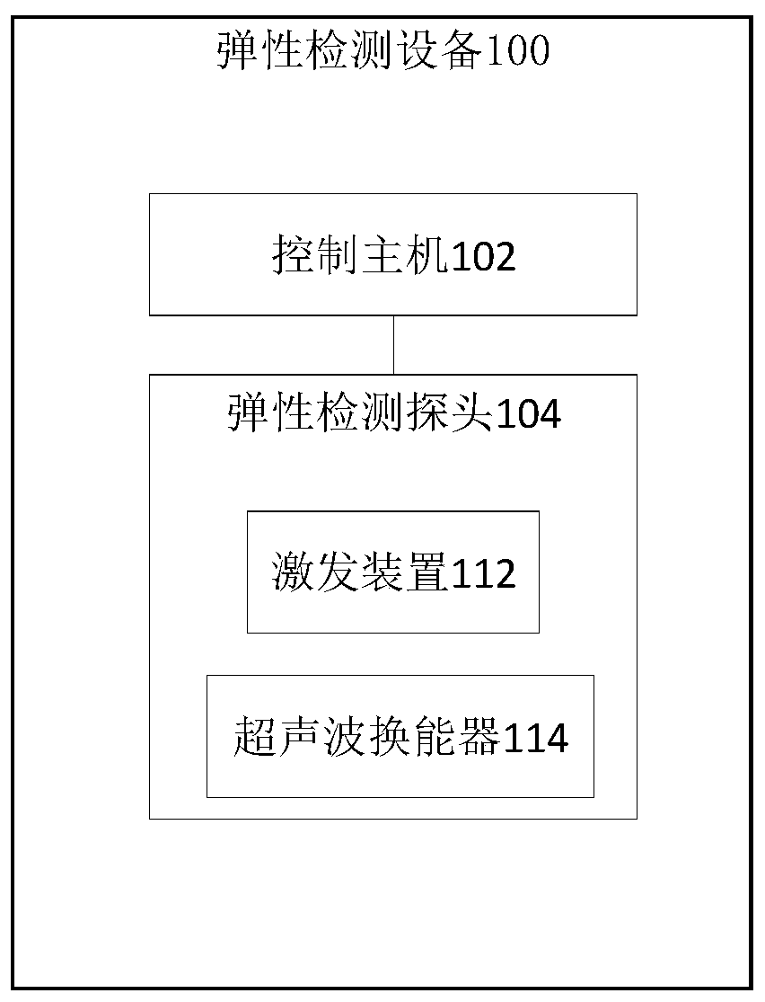
[0034] In an embodiment of the present invention, figure 1 is the structural frame of the tissue elasticity detection device according to the embodiment of the present invention Figure 1 ,Such as figure 1 As shown, the tissue elasticity detection device 100 includes a control host 102 and an elasticity detection probe 104; wherein, figure 2 is the structural frame of the tissue elasticity detection device according to the embodiment of the present invention Figure II ,Such as figure 2 As shown, the elasticity detection probe 104 includes an excitation device 112 an...


PUM


Abstract

The invention discloses a method and equipment for detecting tissue elasticity. According to the method, a first ultrasonic signal is transmitted to a tissue in a detection area; at least one imagingline of the first ultrasonic signal is tracked; a movement state of each imaging line is determined according to the imaging line of a plurality of time points; and a position of an imaging line whosemovement state meets preset conditions is selected, and tissue elasticity detection is carried out. The problem that a movement state of a tissue in an area to be detected affects accuracy of elasticity detection is solved, and the accuracy of elasticity detection for the tissue in the detection area is improved.

Description

technical field [0001] The present application relates to the technical field of medical imaging, in particular to a tissue elasticity detection method and equipment. Background technique [0002] The development of various chronic diseases, such as viral hepatitis (hepatitis A, hepatitis B, hepatitis C, etc.), will be accompanied by fibrosis of damaged tissues, and the process of tissue fibrosis will be accompanied by changes in tissue elasticity. Therefore, tissue elasticity information is a parameter that can be used to diagnose the degree of tissue fibrosis. [0003] Transient Elastography (abbreviated as TE) is a technique for quantitatively detecting tissue elastic modulus, which can comprehensively reflect the degree of tissue fibrosis by detecting liver stiffness measurement (abbreviated as LSM). [0004] However, the transient elastography technique cannot obtain the tissue structure information of the detection area, especially the two-dimensional structure inform...

Claims


Application Information

Patent Timeline
Patent Type & Authority Applications(China)
IPC IPC(8): A61B8/08A61B8/00
CPCA61B8/08A61B8/485A61B8/5207A61B8/5276A61B8/54G01S7/52042G01S15/8915A61B8/4488
Inventor 何琼邵金华孙锦段后利
Owner WUXI HISKY MEDICAL TECH
Why Eureka
Try Eureka