
A nasal cavity filling air bag device for transnasal neuroendoscopic surgery

An air bag and nerve technology, applied in the field of medical equipment, can solve the problems of nasal mucosa damage, narrowed surgical field of view, unfavorable surgery, etc., and achieve the effect of ensuring normal operation

Active Publication Date: 2020-07-31
PEKING UNION MEDICAL COLLEGE HOSPITAL CHINESE ACAD OF MEDICAL SCI
View PDF8 Cites 0 Cited by
  • Summary
  • Abstract
  • Description
  • Claims
  • Application Information

AI Technical Summary

Problems solved by technology

For a long time, due to the rich blood vessels in the nasal cavity, the bleeding and oozing of this channel often lead to poor surgical field of view, which requires repeated washing or even continuous removal of the endoscopic lens to clean it; Lead to re-injury of the nasal mucosa, resulting in increased bleeding; clinically, some units currently use adrenaline cotton to push and pack the nasal mucosa to create a channel, so that the endoscopic lens can enter and exit conveniently, and the nasal mucosa vasoconstriction achieves the purpose of hemostasis; But the disadvantages are also obvious. Absorption of adrenaline by the nasal mucosa will lead to an increase in blood pressure during the operation, resulting in unpredictable postoperative complications, such as intracranial hemorrhage; some units use nasal dilators, but the nasal dilators cannot effectively compress the middle turbinate and the nasal mucosa of the inferior turbinate, resulting in blood still polluting the surgical field of view along the nasal dilator, and the nasal dilator cannot move after being fixed, which limits the mobility of the endoscopic lens and narrows the surgical field of view, which is not conducive to better surgery Can not better meet the needs of the operation

Method used


Image

  • A nasal cavity filling air bag device for transnasal neuroendoscopic surgery
  • A nasal cavity filling air bag device for transnasal neuroendoscopic surgery
  • A nasal cavity filling air bag device for transnasal neuroendoscopic surgery

Examples


Embodiment 1

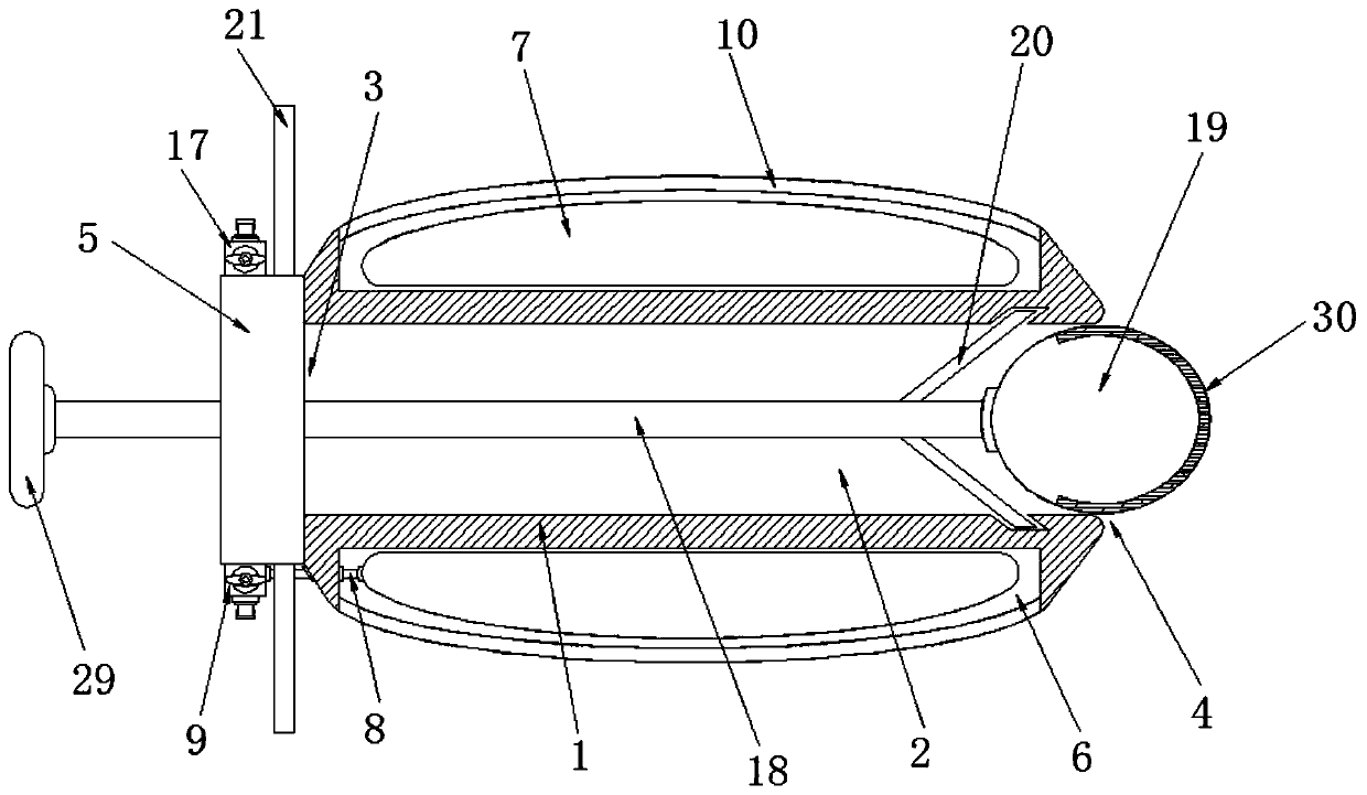
[0030] Example one, such as Figure 1-5 As shown, a nasal cavity filling balloon device for transnasal neuroendoscopic surgery according to an embodiment of the present invention includes a nasal cavity tube 1, and an endoscopic channel 2 is opened inside the nasal cavity tube 1, and the endoscopic channel 2 The two sides of the nasal cavity are respectively provided with an opening 1 3 and an opening 2 4, and the nasal cavity tube 1 is located on one side of the opening 2 4 and has an arc-shaped edge structure. A connecting ring 5 is installed on one side of the opening two 4, an airbag placement cavity 6 is opened on the outer side of the nasal cavity tube 1 away from the endoscope channel 2, and an airbag 7 is installed in the airbag placement cavity 6 through The interaction between the nasal cavity tube 1 and the inflatable bag 7 can facilitate the entry and exit of the endoscopic lens during the operation and facilitate better nasal neuroendoscopic surgery. The inflatable...

Embodiment 2

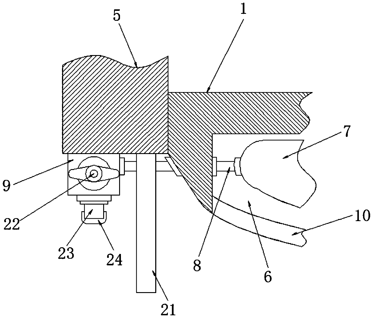
[0031] Example two, such as figure 1 , 2 As shown, a valve switch 22 is installed on the connector 9; an inflation head 23 is installed on the bottom of the connector 9, and a sealing cover 24 is installed on the inflation head 23; The sealing cap 24, the inflation head 23 is connected with the external inflation device, and then the valve switch 22 is turned on, the inflation bag 7 is inflated through the inflation tube 8, and the valve is closed after the inflation is completed Switch 22, the valve switch 22 can ensure that the inflatable bag 7 will not leak air, and ensure that it can be used stably; while the sealing cover 24 protects the structure before use to ensure use The structure of the front device can maintain good stability, so that it can be used better.

Embodiment 3

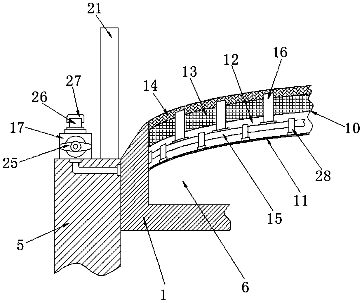
[0032] Example three, such as figure 1 , 3 As shown, a valve switch 25 is installed on the second connector 17; a pipe joint 26 is installed on the top of the connector two 17, and a sealing cover 27 is installed on the pipe joint 26; In the second sealing cap 27, the hemostatic drug syringe is connected to the pipe joint 26, and then the valve switch 25 is turned on, and the hemostatic drug is delivered to the tube through the interaction of the catheter one 15 and the catheter two 16. The inside of the aseptic cotton layer 14, so that the aseptic cotton layer 14 can stop bleeding the blood vessels in the inner lining of the nasal cavity; and the sealing cover 27 protects the structure before use to ensure that the device is protected before use. The structure can maintain good stability, so that it can be used better.


PUM


Abstract

The invention discloses a nasal cavity filling airbag device for transnasal endoscopic neurosurgery. The nasal cavity filling airbag device includes a nasal cavity tube, an endoscope channel is formedin the nasal cavity tube, and an opening I and an opening II are formed in the two side edges of the endoscope channel correspondingly; the side edge, located on the opening II, of the nasal cavity tube is of a curved edge structure, and a connection ring is installed at the side edge, located at the opening I and away from the opening II, of the nasal cavity tube; an airbag placement cavity is formed in the outer side, away from the endoscope channel, of the nasal cavity tube, and an inflation bag is installed in the airbag placement cavity; and an inflation tube is installed at the side edge, close to the opening I, of the inflation bag, the end, away from the inflation bag, of the inflation tube penetrates through the side wall of the nasal cavity tube and extends below the connectionring, and a connection head I is installed below the connection ring. The nasal cavity filling airbag device has the beneficial effect that the transnasal endoscopic neurosurgery can be better assisted, so that the needs during surgery can be better met.

Description

Technical field [0001] The present invention relates to the technical field of medical appliances, in particular to a nasal cavity filling airbag device used for transnasal neuroendoscopic surgery. Background technique [0002] Currently, transnasal neuroendoscopy is used to treat pituitary tumors, skull base tumors and even aneurysms. The nasal cavity as a neuroendoscopy channel is the necessary channel for this operation. For a long time, due to the rich blood vessels in the nasal cavity, the bleeding and oozing of the channel often lead to the deterioration of the surgical field. It is necessary to repeatedly wash and even continuously remove the endoscopic lens and wipe it; it is extremely easy to repeatedly enter and exit the endoscopic lens. Causes the nasal mucosa to be damaged again, which makes bleeding increase. In clinical practice, some units currently use adrenaline cotton to push and compress the nasal mucosa to create a passage, so that the endoscopic lens can be e...

Claims


Application Information

Patent Timeline
Patent Type & Authority Patents(China)
IPC IPC(8): A61B17/12A61B17/24
CPCA61B17/1204A61B17/12136A61B17/24A61B2017/12004
Inventor 姚勇王志成朱建宇李晓旭孙旭郑光耀张毅
Owner PEKING UNION MEDICAL COLLEGE HOSPITAL CHINESE ACAD OF MEDICAL SCI
Learn More