
Peripherally inserted central catheter positioning algorithm based on intracardiac electrogram

A central venous catheter and positioning algorithm technology, applied in catheters, medical science, sensors, etc., can solve problems such as inability to detect catheters and catheter tip position lag in time

Inactive Publication Date: 2020-01-17
SHANDONG BRANDEN MEDICAL DEVICE
View PDF6 Cites 0 Cited by
  • Summary
  • Abstract
  • Description
  • Claims
  • Application Information

AI Technical Summary

Problems solved by technology

However, this method has its disadvantages: judging the position of the catheter tip is still lagging, and usually the abnormal position of the catheter cannot be found in time. For patients with catheter abnormality, it is often necessary to readjust the catheter and take X-rays again for positioning

Method used


Image

  • Peripherally inserted central catheter positioning algorithm based on intracardiac electrogram
  • Peripherally inserted central catheter positioning algorithm based on intracardiac electrogram
  • Peripherally inserted central catheter positioning algorithm based on intracardiac electrogram

Examples


Embodiment 1

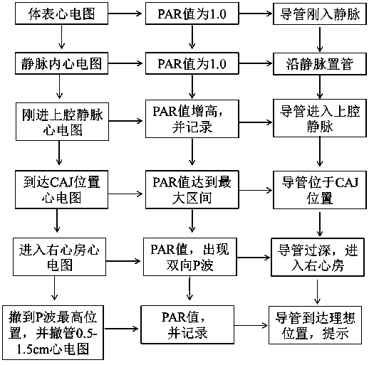
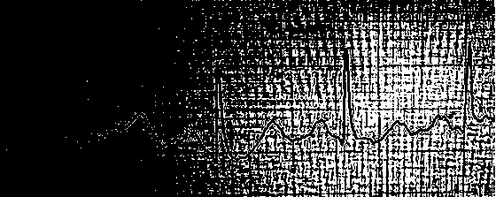
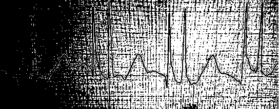
[0031] figure 1 It is a structural diagram of an algorithm for establishing correspondence between the intracavity electrocardiogram and the position of the catheter tip. When the catheter tip is in a peripheral vein (axillary vein, subclavian vein, brachiocephalic vein), an ECG is obtained, such as figure 2 , and measure the P wave amplitude of the electrocardiogram at this time as P1. There is no significant difference between the P wave amplitude of the electrocardiogram at this position and the body surface electrocardiogram, and the PAR value is about 1.0. When the catheter was advanced in the vein, but the catheter tip did not enter the superior vena cava, there was no change in P wave amplitude, and the PAR value was approximately 1.0. However, when the tip of the catheter just entered the superior vena cava, the P wave amplitude suddenly increased significantly. At this time, the P wave amplitude of the electrocardiogram was P2, and the PAR value also increased signi...

Embodiment 2

[0034] Image 6It is a structural diagram of an algorithm for establishing correspondence between the intracavity electrocardiogram and the position of the catheter tip. When the catheter tip is located in the peripheral vein (axillary vein, subclavian vein, brachiocephalic vein), the electrocardiogram is measured, and the ratio of P wave to QRS wave amplitude of the electrocardiogram at this time is measured, and the PQAP value is recorded. When the catheter was advanced intravenously but the catheter tip did not enter the superior vena cava, there was no change in P wave amplitude and no change in PQAP values. When the catheter tip just entered the superior vena cava, the P wave amplitude suddenly increased significantly, and the PQAP value also increased significantly. As the catheter tip advances in the superior vena cava, the recorded PQAP value gradually increases; when the catheter tip is located at the junction of the superior vena cava and the right atrium, the P wav...

Embodiment 3

[0037] Confirming the position of the tip of the catheter according to a parameter may cause deviations in insertion due to individual differences. PAR and PQAP values ​​can be used as dual parameters to guide catheter placement. When both the PAR value and the PQAP value enter the ideal range, it means that the tip of the catheter is placed in the ideal area to further improve the accuracy rate.


PUM


Abstract

The invention relates to a peripherally inserted central catheter positioning algorithm based on intracardiac electrogram, and belongs to the field of medical apparatuses. The peripherally inserted central catheter positioning algorithm based on intracardiac electrogram comprises the steps of firstly, performing checking to obtain the electrocardiograph of the head of a peripherally inserted central catheter at different positions, calculating the ratio of P wave amplitude of the electrocardiograph at different positions to P wave amplitude of the surface electrocardiogram to obtain PAR value;and calculating the amplitude ratio of P waves to QRS waves of the electrocardiograph at different positions to obtain PQAP, so as to establish the relationship of the position of the head of the peripherally inserted central catheter to the PAR value or the PQAP value. Through data statistics, a method for expressing the position of the head of the peripherally inserted central catheter with PARvalue or the PQAP value is finally established. In the process of placing the peripherally inserted central catheter, equipment can measure the electrocardiograph, can display the PAR value or the PQAP value and the expectation position of the corresponding peripherally inserted central catheter, can prompt the position of the peripherally inserted central catheter in a real time manner, and canperform voice prompt that the peripherally inserted central catheter achieves the expectation position. The electrocardiograph is directly changed into the PAR value or the PQAP value and the positionof the head of the corresponding peripherally inserted central catheter, medical personnel can directly read the position of the tip of the peripherally inserted central catheter, and positioning ofthe peripherally inserted central catheter can be quickly and conveniently completed.

Description

technical field [0001] The invention belongs to the field of medical devices, and more specifically designs a positioning algorithm for a transperipheral central venous catheter based on intracavitary electrocardiography. Background technique [0002] According to statistics from the National Cancer Center, cancer incidence in China accounted for about 22% of the world's total in 2017, with a standardized incidence rate of 174.0 / 100,000 and a world average of 182.3 / 100,000. Chemotherapy is an important means of medical oncology to treat malignant tumors, and the commonly used route of administration is repeated superficial venous puncture. Because chemotherapy drugs stimulate blood vessels, it will inevitably cause vascular damage. [0003] Peripherally Inserted Central Catheter (PICC), as the lifeline of patient treatment, plays an important role in promoting patient recovery. PICC catheters provide a safe and long-term vascular access for patients who need chemotherapy, ...

Claims


Application Information

Patent Timeline
IPC IPC(8): A61M25/01A61B5/0402
CPCA61M25/01A61B5/318
Inventor 张海军刘光支树迪韩真真李宜玮侯文博周超尹玉霞鲁手涛袁坤山
Owner SHANDONG BRANDEN MEDICAL DEVICE
Learn More