A scanning imaging system for interventional treatment

A scanning imaging, interventional technology, applied in image enhancement, image analysis, medical image and other directions, can solve the problems of difficult image collection and poor imaging effect, to avoid format incompatibility and ensure the effect of unified processing

Inactive Publication Date: 2020-07-10
张军 +3
View PDF6 Cites 0 Cited by
  • Summary
  • Abstract
  • Description
  • Claims
  • Application Information

AI Technical Summary

Problems solved by technology

[0005] In order to solve the problems of difficult image collection and poor imaging effect during interventional treatment in the current medical scanning imaging process, the present invention claims a scanning imaging system for interventional treatment, which is characterized in that it includes:

Method used


Image

  • A scanning imaging system for interventional treatment
  • A scanning imaging system for interventional treatment
  • A scanning imaging system for interventional treatment

Examples


Embodiment Construction

[0014] The following will clearly and completely describe the technical solutions in the embodiments of the present invention with reference to the accompanying drawings in the embodiments of the present invention. Obviously, the described embodiments are only some, not all, embodiments of the present invention. Based on the embodiments of the present invention, all other embodiments obtained by persons of ordinary skill in the art without creative efforts fall within the protection scope of the present invention.

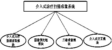
[0015] Refer to attached figure 1 , the present invention claims a module structure diagram of a scanning imaging system for interventional therapy.

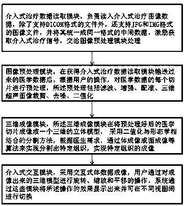
[0016] The present invention claims a scanning imaging system for interventional treatment, which is characterized in that it includes, referring to the attached figure 2 A working flow chart of each module of the scanning imaging system for interventional treatment of the present invention:

[0017] The intervent...


PUM


Abstract

According to a scanning imaging system for interventional therapy, an interventional therapy data reading module is used for reading the interventional therapy data. The system comprises an image preprocessing module. After medical data transmitted by the interventional therapy data reading module is obtained, according to operation of a user, each slice of the medical data is preprocessed, specific tissue is segmented through algorithms such as volume imaging or surface imaging, imaging of the specific tissue is achieved, and finally the system displays the operation effect through the modules and can be switched between different views. Effective collection and format unification processing of equipment images in the interventional medical process are effectively guaranteed, incompatibility of formats of different medical images is avoided, the interventional images are effectively processed and stored, the imaging effect of related scanning images is guaranteed, and the query analysis efficiency is improved.

Description

technical field [0001] The present application relates to the technical field of medical image segmentation, in particular to a method and device for segmenting three-dimensional medical images. Background technique [0002] Medical image segmentation is the basis for subsequent processing such as 3D visualization of normal and diseased tissues, surgical simulation, and graphic-guided surgery. The accuracy of segmentation is crucial for doctors to judge the true condition of a patient's disease and make a corresponding diagnosis plan. [0003] Many ultrasound imaging systems output video at a fixed frame rate. For example, images are output at 30 Hz or 60 Hz to simplify conversion to the NTSC standard video signal required by videocassette recorders. Matching the video frame rate to that used in typical applications simplifies the recording of ultrasound scans. Off-the-shelf components can also be used to generate video in ultrasound systems. [0004] Since current medica...

Claims


Application Information

Patent Timeline
Patent Type & Authority Patents(China)
IPC IPC(8): G06T17/00G06T19/20G06T7/11G06T7/155G16H30/20G16H30/40
CPCG06T7/11G06T7/155G06T17/00G06T19/20G06T2200/08G06T2207/10136G06T2207/30004G06T2219/2016G16H30/20G16H30/40
Inventor 张军宋平陈瑞萍魏红梅
Owner 张军
Why Eureka
Try Eureka