Method and device for improving stability of examined part in medical image examination process

A technology of medical imaging and inspection process, which is applied in medical science, patient positioning for diagnosis, and equipment for radiological diagnosis, etc. Clamping and other problems, to achieve the effect of firm fixation, high safety and convenient operation

Active Publication Date: 2020-03-27
WEST CHINA HOSPITAL SICHUAN UNIV
View PDF8 Cites 2 Cited by
  • Summary
  • Abstract
  • Description
  • Claims
  • Application Information

AI Technical Summary

Problems solved by technology

However, this method has the following disadvantages: 1. For patients with different body shapes, sponge wedges of different shapes need to be prepared, which is inconvenient to use; It is easy to lose the fixing effect on the tested part, resulting in the failure of the test; 3. The sponge wedge cannot be evenly pressed on the tested part, which is likely to cause discomfort to the patient and stimulate the patient to actively adjust the body position, resulting in the failure of the test

Method used


Image

  • Method and device for improving stability of examined part in medical image examination process
  • Method and device for improving stability of examined part in medical image examination process
  • Method and device for improving stability of examined part in medical image examination process

Examples


Embodiment 1

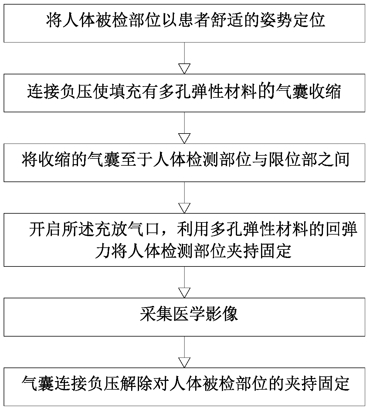
[0041] Embodiment 1: A method for improving the stability of the inspected part in the process of medical image inspection.

[0042] Such as figure 1 As shown, this embodiment provides a method for improving the stability of the inspected part in the process of medical image examination, by fixing the inspected part of the human body in an adaptive fixation manner; and then collecting the medical image. The adaptive fixation method refers to the use of deformable substances to fit and squeeze the fixed object based on the shape of the fixed object, so as to form a contact surface specific to the contour of each fixed object, so the pressure is more uniform , higher comfort, and more secure fixation. Specifically, the method of adaptive fixation includes: step 1, positioning the human body to be tested in a comfortable posture for the patient; step 2, using a device capable of generating adaptive deformation to clamp and fix the human body to be tested. The final clamping and...

Embodiment 2

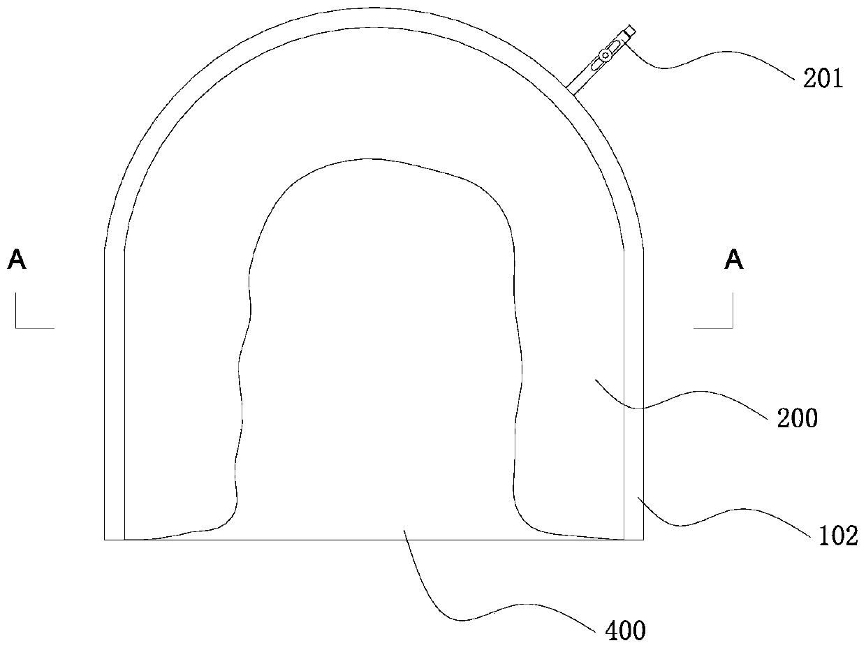
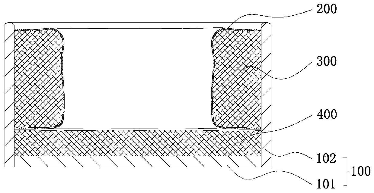
[0047] Embodiment 2: A device for improving head stability during medical imaging examination.

[0048] Such as figure 2 and image 3 As shown, the device includes: a support shell 100, an airbag 200 and a porous elastic material 300; the support shell 100 includes a base plate 101 and a U-shaped plate 102 vertically installed on the upper surface of the base plate 101, and the support shell 100 can communicate with medical imaging equipment through the base plate 101 Connect to play the role of connection and fixation; the airbag 200 is U-shaped, the outer surface of the airbag 200 is connected with the inner surface of the U-shaped plate 102, and the expansion of the airbag 200 occupies the inner space of the supporting shell 100, which can clamp the part to be tested. The airbag 200 is provided with an inflation and deflation port 201 to realize the discharge and filling of gas inside the airbag 200; the porous elastic material 300 is filled in the airbag 200, and the por...

Embodiment 3

[0051] Embodiment 3: A device for improving trunk stability during medical imaging examination.

[0052] Such as Figure 5 and Figure 6 As shown, compared with Embodiment 2, the main difference lies in the change of the shape of the supporting shell 100 and the arrangement of the airbag 200 . The supporting shell 100 includes a base plate 101 and two side plates 103 vertically installed on the upper surface of the base plate 101 , and airbags 200 are respectively installed on opposite surfaces of the two side plates 103 .

[0053] Such as Figure 7 As shown, when in use, the patient raises his arms and lays his body on his back. The torso is located between the two side panels 103. After the inflation and deflation port 201 is opened, the airbags 200 bulge from the left and right sides and fit the left and right sides of the patient's torso. achieve a fixed effect.


PUM


Abstract

The invention discloses a method and device for improving the stability of an examined part in the medical imaging examination process, and belongs to the technical field of medical imaging equipment.According to the method, after the body position of a patient is placed, the examined part of the human body is fixed in an adaptive fixing mode; the medical image is acquired, so as to be suitable for patients with different figures and body types and equipment with different models, and the device is simple to operate, firmer to fix and higher in comfort level. Due to the fact that the air bagwraps the porous elastic material, free circulation of air in the porous elastic material and the atmosphere is limited, after the air bag is connected with negative pressure, the air bag can flattenthe porous elastic material under the pressure of the atmospheric pressure, and the elastic potential energy of the porous elastic material is increased. After the air bag is communicated with the atmosphere, the elastic potential energy of the porous elastic material is gradually released to support the air bag, so that a high-pressure air source is not needed; high-pressure inflation is not needed after the air bag is placed in place, and operation is convenient; no air leakage or explosion risk exists in the image inspection process, and the safety coefficient is very high.

Description

technical field [0001] The invention belongs to the technical field of medical imaging equipment, and in particular relates to a method and a device for improving the stability of an inspected part in the medical imaging inspection process. Background technique [0002] Medical imaging refers to the technology and processing process of obtaining internal tissue images of the human body or a certain part of the human body in a non-invasive manner for medical treatment or medical research. With the continuous advancement of science and technology, the images obtained by people have developed from simple two-dimensional to three-dimensional, from morphology to functionality, from qualitative to quantitative. [0003] Conventional X-ray imaging only needs to collect data once, which can be completed in an instant, but the patient still needs to remain still. Both CT (Computed Tomography) and MRI (Magnetic Resonance Imaging) require continuous examination of the human body withi...

Claims


Application Information

Patent Timeline
Patent Type & Authority Applications(China)
IPC IPC(8): A61B6/04A61B5/055
CPCA61B5/055A61B6/04A61B6/0407A61B6/0485
Inventor 夏春潮李玉明曾令明邓丽萍李磊赵飞
Owner WEST CHINA HOSPITAL SICHUAN UNIV
Why Eureka
Try Eureka