Fundus image micro-hemangioma automatic detection method and storage equipment

A technology for fundus images and microangiomas, applied in image enhancement, image analysis, image data processing, etc., can solve the problems of complex calculations and low efficiency of analysis methods for fundus images of microangiomas, achieve low contrast, small targets, reduce diagnosis and Effect

Active Publication Date: 2020-03-27
FUZHOU YIYING HEALTH TECH CO LTD
View PDF5 Cites 2 Cited by
  • Summary
  • Abstract
  • Description
  • Claims
  • Application Information

AI Technical Summary

Problems solved by technology

[0006] To this end, it is necessary to provide an automatic detection method for microvascular tumors in fundus images, which is used to solve the problems of complex calculations in detecting microvascular tumors in fundus images in the prior art, requiring a large amount of data for training, and low efficiency of analysis methods

Method used


Image

  • Fundus image micro-hemangioma automatic detection method and storage equipment
  • Fundus image micro-hemangioma automatic detection method and storage equipment
  • Fundus image micro-hemangioma automatic detection method and storage equipment

Examples


Embodiment Construction

[0023] In order to explain in detail the technical content, structural features, achieved goals and effects of the technical solution, the following will be described in detail in conjunction with specific embodiments and accompanying drawings.

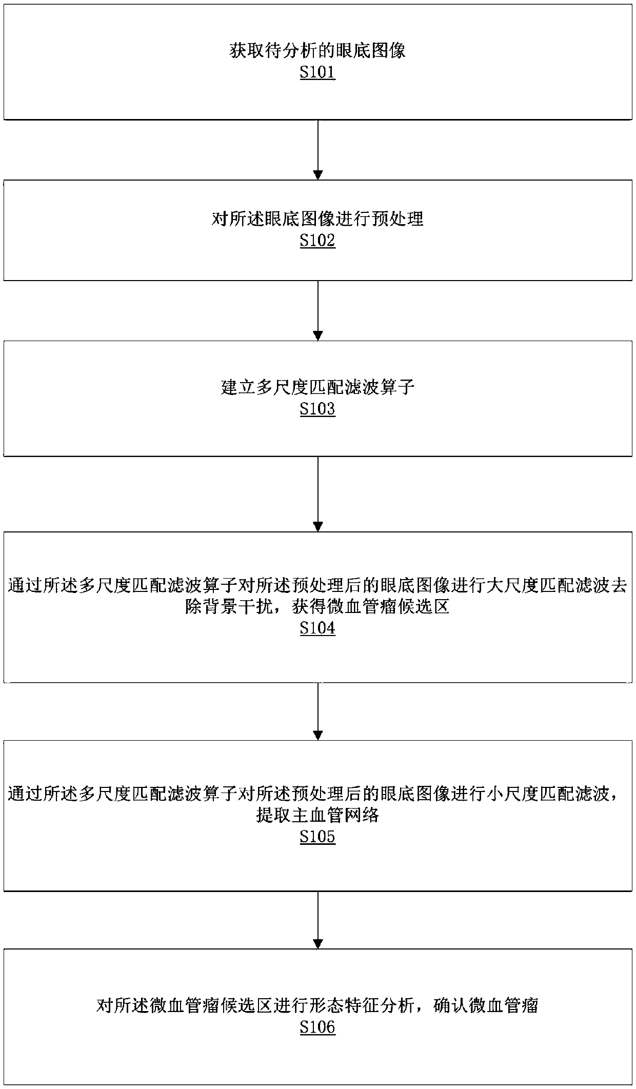
[0024] see figure 1 , in this embodiment, a method for automatic detection of microvascular tumors in fundus images can be applied to a storage device. In this embodiment, a storage device can be a smart phone, a tablet computer, a desktop PC, a notebook computer, a PDA etc.

[0025] In this embodiment, a specific implementation of a method for automatic detection of microvascular tumors in fundus images is as follows:

[0026] Step S101: Obtain a fundus image to be analyzed. The following methods can be adopted: collect the fundus image of the subject through the fundus camera, and then upload the fundus image of the corresponding subject to the storage device for processing, or directly input the fundus image of the subject, or ob...


PUM


Abstract

The invention relates to the field of fundus image processing, in particular to a fundus image micro-hemangioma automatic detection method and storage equipment. The fundus image micro-hemangioma automatic detection method comprises the steps of acquiring a fundus image to be analyzed; preprocessing the fundus image; establishing a multi-scale matched filtering operator; performing large-scale matched filtering on the preprocessed fundus image to remove background interference to obtain a micro-hemangioma candidate region; performing small-scale matched filtering on the preprocessed fundus image, and extracting a main blood vessel network; and performing morphological characteristic analysis on the micro-hemangioma candidate region to confirm the micro-hemangioma. The whole process does not need a large amount of data to carry out deep learning training, a doctor is simply, effectively and rapidly helped to exclude fundus photos without hemangioma or diabetic patients without DR, and the workload of further diagnosis and large-scale DR screening traditional Chinese medicine doctor interpretation grading is greatly reduced.

Description

technical field [0001] The invention relates to the field of fundus image processing, in particular to a method for automatically detecting microvascular tumors in fundus images and a storage device. Background technique [0002] According to the World Health Organization, 347 million people worldwide are diagnosed with diabetes, and this number is expected to exceed 640 million by 2040. Evidence-based medical research has proved that high blood sugar, high blood pressure, high blood lipids and the course of diabetes are important risk factors for the occurrence of diabetic retinopathy (English abbreviation "DR"). Therefore, strict control of blood sugar, blood lipids, blood pressure and other risk factors, as well as fundus screening, can significantly reduce the risk of diabetic retinopathy in diabetic patients. [0003] The experience of various countries has shown that: "telemedicine is an effective way to carry out DR screening and improve the screening rate and patien...

Claims


Application Information

Patent Timeline
Patent Type & Authority Applications(China)
IPC IPC(8): G06T7/00G06T7/11G06T7/136G06T7/62
CPCG06T7/0012G06T7/11G06T7/136G06T7/62G06T2207/10024G06T2207/20081G06T2207/30041G06T2207/30096
Inventor 余轮曹新容王丽纳林嘉雯
Owner FUZHOU YIYING HEALTH TECH CO LTD
Why Eureka
Try Eureka