Method for predicting HER2 gene amplification state of breast cancer HER2 immunohistochemical 2 + samples
A technology of immunohistochemistry and gene amplification, which is applied in the fields of biochemical equipment and methods, measuring devices, and microbial determination/inspection. problem, to achieve the effect of improving detection efficiency, narrowing the scope of pointers, and saving medical costs
Pending Publication Date: 2020-04-10
XIANGYA HOSPITAL CENT SOUTH UNIV
View PDF2 Cites 0 Cited by
- Summary
- Abstract
- Description
- Claims
- Application Information
AI Technical Summary
Problems solved by technology
[0004] HER2-FISH detection of HER2 gene amplification status has the advantages of high accuracy and good specificity, but the detection process is relatively complicated, the experimental conditions are relatively high, it is not easy to carry out in county-level hospitals, and the detection cost is relatively high (for example, Zhongnan HER2-FISH test at University Xiangya Hospital costs 2250 RMB, while immunohistochemical test is only 150 RMB per item)
Some patients may give up testing due to economic reasons, resulting in missed opportunities for targeted therapy
Method used
View moreImage
Smart ImageViewing Examples

Examples
Embodiment 1
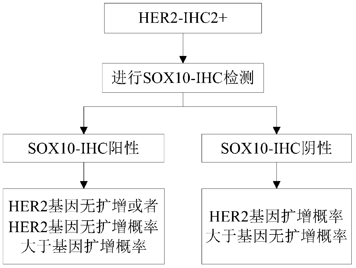
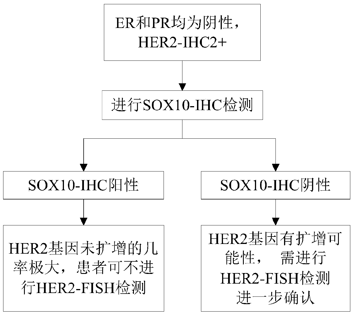
[0030] see figure 2 , this embodiment is a method for predicting the state of HER2 gene amplification in breast cancer HER2 immunohistochemical 2+ samples, comprising the following steps:
[0031] The samples used in this example are paraffin-embedded samples of isolated tissues obtained after breast lesion puncture or surgery. Each paraffin-embedded sample can be subjected to multiple immunohistochemical detection and FISH detection, which is highly reproducible .
Login to View More PUM
 Abstract
The invention discloses a method for predicting an HER2 gene amplification state in a breast cancer HER2 immunohistochemical 2 + samples. The method comprises the following steps: SOX10 immunohistochemical detection is carried out on a breast cancer HER2 immunohistochemical 2 + sample, and samples with the positive SOX10 immunohistochemical detection result are determined as no amplification of HER2 gene or greater probability of no amplification of HER2 gene than probability of gene amplification. According to the invention, blindness of the FISH detection on all HER2 immunohistochemical 2 +samples is avoided to a certain extent, the clinical pointer range of HER2-FISH detection is narrowed, and the medical cost is remarkably saved.
Description
technical field [0001] The invention relates to a screening method of gene expression profiles, in particular to a method for predicting the HER2 gene amplification status of breast cancer HER2 immunohistochemical 2+ samples. Background technique [0002] At present, there are two commonly used detection methods for HER2 (Human Epidermal growth factor Receptor-2, human epidermal growth factor receptor-2) in the world, namely, immunohistochemistry (IHC, Immunohistochemistry) and fluorescence in situ hybridization (FISH, Fluorescence). In Situ Hybridization) method. The US Food and Drug Administration (FDA, Food and Drug Administration) and the American Society of Clinical Oncology / American Society of Pathologists (ASCO / CAP, American Society of Clinical Oncology / American Society of Pathologists) recommend using two methods for HER2 status For detection, HER2 immunohistochemical detection should be carried out at the protein level first. The samples with HER2 immunohistochemic...
Claims
Login to View More Application Information
Patent Timeline
 IPC IPC(8): C12Q1/6886C12Q1/6841G01N33/574
CPCC12Q1/6886C12Q1/6841G01N33/57415G01N33/57484
Inventor 王宽松粟诗童杨芝春郭伟周训检戚嘉琳胡祯敏唐萍周建华
Owner XIANGYA HOSPITAL CENT SOUTH UNIV