
Tooth image soft and hard tissue detection method

A detection method and image technology, applied in the computer field, can solve the problems of difficult selection of thresholds, missed detection, low contrast of tooth images, etc.

Active Publication Date: 2020-04-14
苏州喆安医疗科技有限公司
View PDF8 Cites 1 Cited by
  • Summary
  • Abstract
  • Description
  • Claims
  • Application Information

AI Technical Summary

Problems solved by technology

[0003] Edge-based detection is based on the characteristics of discontinuous gray levels in the soft and hard tissue areas of dental images, using gradient differential operators such as Sobel and Canny to detect the contours of different regions of teeth, but this method is sensitive to noise, and false edges or The case of missed detection at the edge
[0004] Region-based detection includes threshold segmentation, region growth, region splitting and merging. In threshold segmentation, due to the characteristics of low contrast and uneven gray distribution of tooth images, the selection of threshold becomes very difficult; region growing, region splitting and merging methods The focus is on the regular design, but the splitting process often destroys the edge of dental tissue detection
[0005] In addition to these two, some specific theoretical segmentation detection methods have also been proposed, such as the morphological watershed algorithm. Although this method can effectively segment low-contrast images, over-segmentation often occurs; tooth images based on level sets Segmentation, using the mean and variance of Gaussian fitting energy to describe the local gray intensity of the image, has the advantages of being insensitive to the initial position and stable and unique numerical solution, but the acquisition of prior knowledge is difficult, and the calculation is relatively complicated

Method used


Image

  • Tooth image soft and hard tissue detection method
  • Tooth image soft and hard tissue detection method
  • Tooth image soft and hard tissue detection method

Examples


Embodiment Construction

[0043] The technical solutions in the embodiments of the present invention will be described in detail below in conjunction with the accompanying drawings in the embodiments of the present invention. Obviously, the described embodiments are only some of the embodiments of the present invention, not all of them. Based on the embodiments of the present invention, all other embodiments obtained by persons of ordinary skill in the art without making creative efforts belong to the protection scope of the present invention.

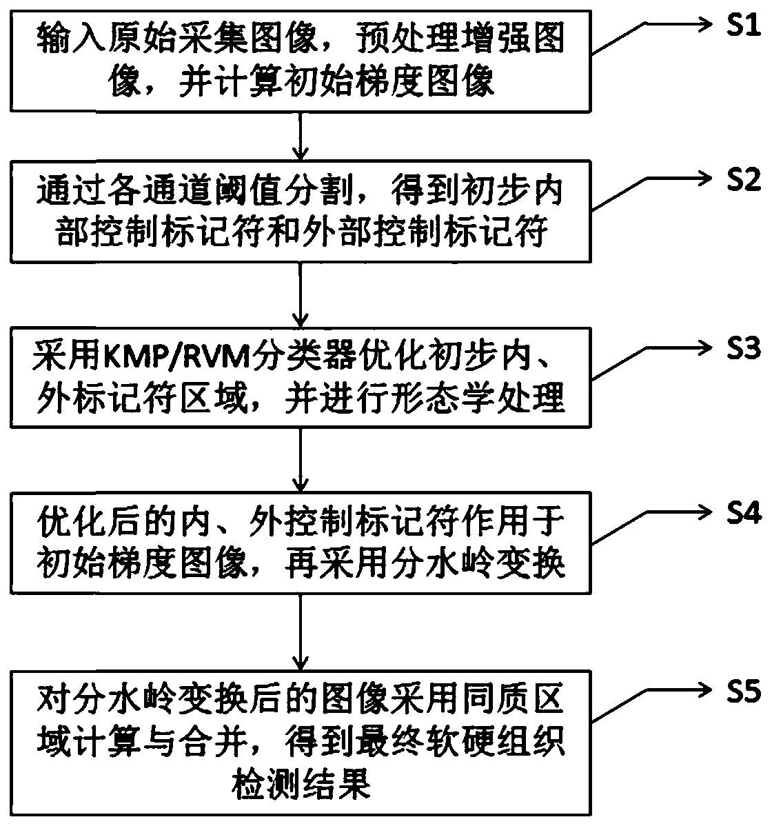
[0044] combine figure 1 Shown, a kind of dental soft and hard tissue detection method comprises:

[0045] S1: Read in the tooth source image obtained from the oral cavity detection equipment, obtain the enhanced image after preprocessing, and calculate its initial gradient image;

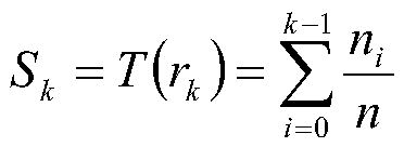
[0046] In this embodiment, ① preprocessing is to use histogram equalization processing to enhance the contrast of the original image; ② the calculation of the initial gradient image...


PUM


Abstract

The invention discloses a tooth image soft and hard tissue detection method. The method comprises the following steps: acquiring an enhanced image of a tooth source image; obtaining an adjusted initial gradient image according to a gradient calculation result and the enhanced image; obtaining a preliminary internal control marker set and a preliminary external control marker set according to a threshold segmentation rule based on each color channel of the tooth source image; obtaining an optimized internal control marker set and an optimized external control marker set according to KMP kernelmatching pursuit or an RVM correlation vector machine algorithm based on the preliminary internal control marker set and the preliminary external control marker set; obtaining a final optimized internal control marker set and a final optimized external control marker set according to a mathematical morphology algorithm; applying the final optimized internal control marker set and the external control marker set on the initial gradient image to obtain a modified gradient image; and finally obtaining images of a soft tissue region and a hard tissue region of the tooth body according to a watershed transformation method and the combination of the homogeneous regions.

Description

technical field [0001] The present application relates to the field of computer technology, in particular to a method for detecting soft and hard tissues of dental images. Background technique [0002] The current detection methods for digital dental images mainly include edge-based detection and region-based detection. [0003] Edge-based detection is based on the characteristics of discontinuous gray levels in the soft and hard tissue areas of dental images, using gradient differential operators such as Sobel and Canny to detect the contours of different regions of teeth, but this method is sensitive to noise, and false edges or The case of missed edge detection. [0004] Region-based detection includes threshold segmentation, region growth, region splitting and merging. In threshold segmentation, due to the characteristics of low contrast and uneven gray distribution of tooth images, the selection of threshold becomes very difficult; region growing, region splitting and ...

Claims


Application Information

Patent Timeline
IPC IPC(8): G06T7/00G06T7/136G06T7/194G06T5/00G06K9/62
CPCG06T7/0012G06T7/136G06T7/194G06T2207/30036G06T2207/20152G06T2207/20036G06F18/241G06T5/90
Inventor 程斌
Owner 苏州喆安医疗科技有限公司
Learn More