
Method for evaluating CAR-T killing activity in vitro

A technology with killing activity and specificity, applied in the field of cell biology, can solve problems such as inability to meet

Inactive Publication Date: 2020-04-21
XUZHOU MEDICAL UNIV
View PDF8 Cites 6 Cited by
  • Summary
  • Abstract
  • Description
  • Claims
  • Application Information

AI Technical Summary

Problems solved by technology

At present, RTCA is used in cell proliferation detection, compound and cytokine-mediated cytotoxicity, cell migration and invasion detection, cell quality control, cell adhesion and extension detection, receptor-mediated signaling pathway detection, cell-cell interaction (co-culture), virus-mediated cytopathic (viral CPE) detection, NK cell-mediated cell killing effect detection, drug screening application, drug cardiomyocyte toxicity detection, etc., but no research has yet applied it to CAR -T in vitro killing activity detection, the reason is that the main therapeutic target of CAR-T is hematological tumors expressing a certain target antigen, and hematological tumor cells are mostly suspension cells, which cannot meet the requirement that RTCA can only detect adherent cell signals

Method used


Image

  • Method for evaluating CAR-T killing activity in vitro
  • Method for evaluating CAR-T killing activity in vitro
  • Method for evaluating CAR-T killing activity in vitro

Examples


Embodiment 1

[0047] 1. Experimental materials:

[0048] SKBR3 cells; CD19 virus LV-CD19 (26043-1) (Jikai Gene); infection enhancer polybrene (Jikai Gene); puromycin (vicmed); PE-CD19 flow cytometry antibody (Sanjian Bio); DMEM medium ( Hyclone) (containing 10% FBS);

[0049] Instrumentation: Flow Cytometry (BD); RTCA (Agilent Bio); Microscope (Olympus).

[0050] 2. Experimental method:

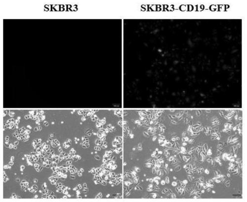
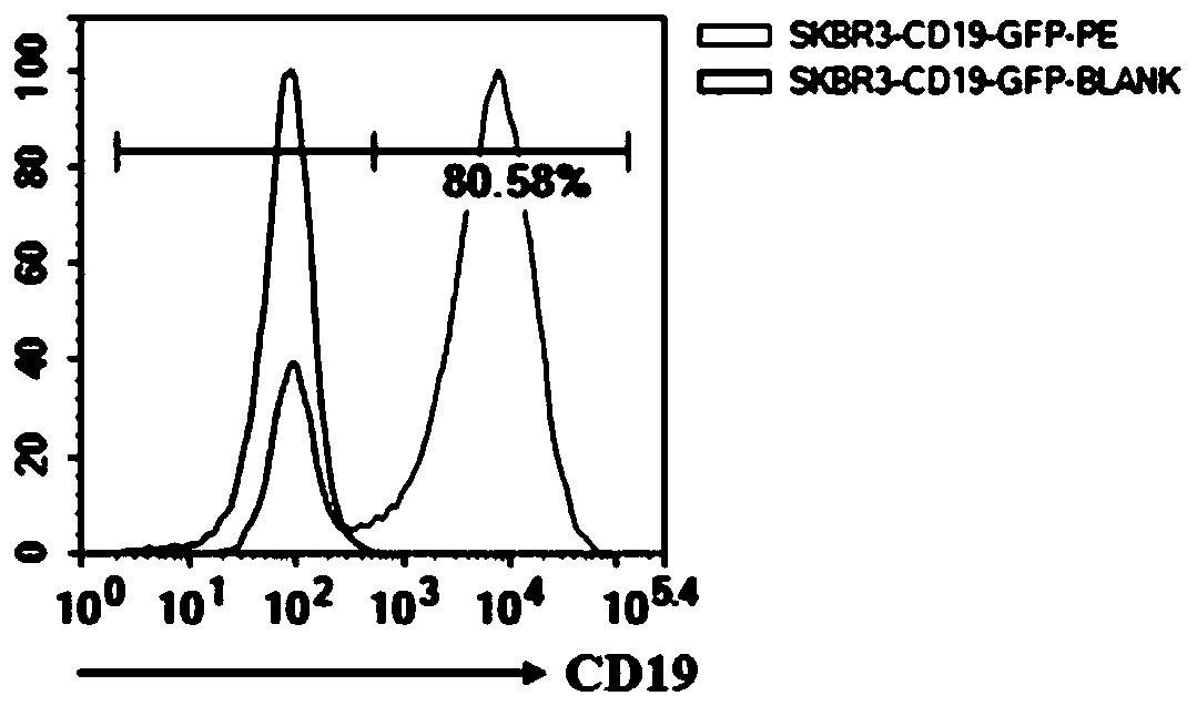
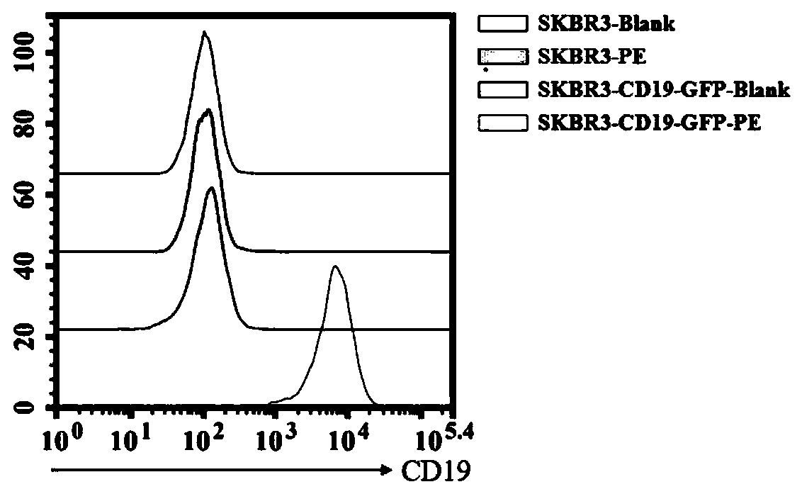
[0051] 1. CD19 virus infected SKBR3 cells.

[0052] The optimal infection conditions (including the amount of inoculated cells, the infection reagent and the time of changing the medium after infection) and the multiplicity of infection (MOI) of the virus to SKBR3 cells were determined by the pre-infection experiment. The specific operation steps are as follows:

[0053] (1) One day before infection, SKBR3 cells in good growth state were prepared with DMEM medium (containing 10% FBS) at a density of 3.75×10 5 cells / ml cell suspension, the cell suspension was 2ml / well (7.5×10 5 Cells / well) were inocul...

Embodiment 2

[0082]1. Experimental materials:

[0083] MDA-MB-231 cells; CD19 virus LV-CD19 (26043-1) (Jikai Gene); infection enhancer polybrene (Jikai Gene); puromycin (vicmed); PE-CD19 flow antibody (Sanjian Biological); DMEM medium (Hyclone) (containing 10% FBS);

[0084] Instrumentation: Flow Cytometry (BD); RTCA (Agilent Bio); Microscope (Olympus).

[0085] 2. Experimental method:

[0086] 1. CD19 virus infected MDA-MB-231 cells.

[0087] The optimal infection conditions (including the amount of inoculated cells, infection reagents, and the time of changing medium after infection) and the multiplicity of infection (MOI) of the virus on MDA-MB-231 cells were determined through the pre-infection experiment. The specific operation steps are as follows:

[0088] (1) One day before infection, the MDA-MB-231 cells in good growth state were prepared with DMEM medium (containing 10% FBS) at a density of 3.75×10 5 cells / ml cell suspension, the cell suspension was 2ml / well (7.5×10 5 Cells...


PUM


Abstract

The invention relates to a method for evaluating CAR-T killing activity in vitro. IN the prior art, the existing CAR-T cell in-vitro killing activity detection method relates to isotope safety, operation is tedious and other problems exist. Based on the problems in the prior art, in order to conveniently and simply realize the detection of in vitro killing activity of CAR-T cells, a cell strain capable of evaluating the CAR-T killing function in vitro is constructed, and a method for detecting the CAR-T in-vitro killing activity through RTCA is provided, wherein the CAR-T cell killing activitycan be efficiently, sensitively and continuously detected in vitro through the method so as to provide the guarantee for the subsequent treatment effect of CAR-T cells.

Description

technical field [0001] The invention relates to the field of cell biology, in particular to a method for evaluating CAR-T killing activity in vitro. Background technique [0002] The in vitro evaluation process of the tumor killing activity of immune cells involves the existence of two types of cells, immune cells and tumor cells. Therefore, traditional cell proliferation activity detection methods (such as MTT, ATP, etc.) cannot be used for direct detection, but different methods should be used. means of marking. Currently used evaluation methods include the following: (1) radioisotope 51 Cr release detection, isotope 51 Cr release is the most classic cytotoxicity detection method for evaluating the killing of tumor cells by CAR-T immune cells. 51 Cr can specifically label target tumor cells, wash and centrifuge to remove excess 51 After Cr, CAR-T immune cells are added for co-cultivation. After the immune cells start the killing program, the target tumor cells are lyse...

Claims


Application Information

Patent Timeline
IPC IPC(8): C12Q1/18C12N5/10
CPCC07K14/70503C12N5/0693G01N33/5011G01N33/505
Inventor 刘丹徐溪悦施明刘庶慈汤安群郑骏年
Owner XUZHOU MEDICAL UNIV
Learn More