
Augmented-reality microscope and augmented-reality method thereof

A technology of augmented reality and microscopy, applied in ophthalmoscopy, medical science, surgery, etc., can solve problems such as observation and diagnosis, safety risks, and increased workload for doctors, so as to improve the effect, achieve separation, and reduce the workload.

Pending Publication Date: 2020-06-23
SHANGHAI MEDIWORKS PRECISION INSTR CO LTD
View PDF3 Cites 0 Cited by
  • Summary
  • Abstract
  • Description
  • Claims
  • Application Information

AI Technical Summary

Problems solved by technology

[0003] The above-mentioned existing technical solutions have the following defects: for the inspection with the output of analysis information and the current state information of the device itself (such as the brightness of the light source, the type of the light source, the current magnification, etc.), the doctor needs to often check the information on the computer screen , unable to observe and diagnose directly from the eyepiece, it is necessary to switch the line of sight back and forth, which increases the workload of the doctor and also brings safety risks

Method used


Image

  • Augmented-reality microscope and augmented-reality method thereof
  • Augmented-reality microscope and augmented-reality method thereof
  • Augmented-reality microscope and augmented-reality method thereof

Examples


Embodiment 1

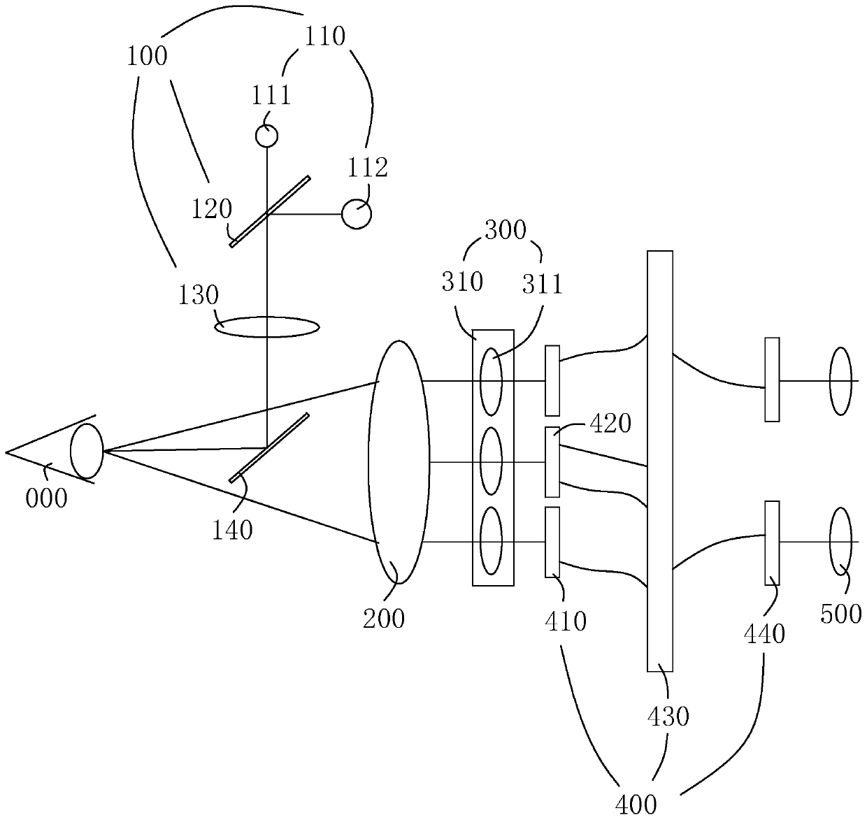
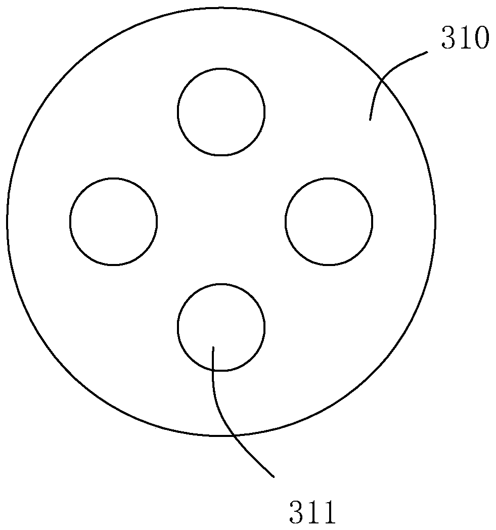
[0056] refer to figure 1 , is a reality-augmented microscope disclosed in the present invention, including a light source module 100 for illuminating a target object 000, an objective lens and a zoom group 200 for receiving reflected light signals reflected by the target object 000, and a zoom group 200 for separating objects passing through the objective lens and The separation module 300 of the reflected optical signal of the variable magnification group 200, and the processing module 400 for analyzing the optical signal and outputting the optical result.

[0057] The light source module 100 includes a working light source 110 that provides at least one kind of light, and can also use a working light source 110 that provides different kinds of light, or a working light source 110 that provides a continuous spectrum. The light of different working light sources 110 passes through the first diphasic color The mirror 120 , the first projection lens 130 and the mirror 140 reach ...

Embodiment 2

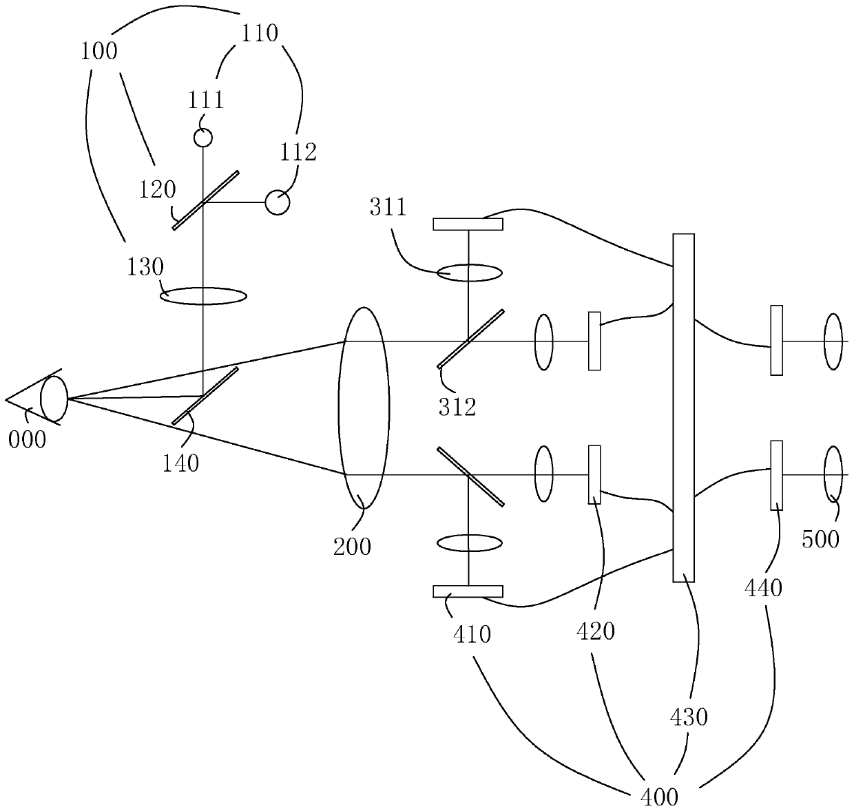
[0063] refer to image 3 , is a reality augmented microscope disclosed in the present invention, and is a reality augmented microscope disclosed in the present invention. The difference from Embodiment 1 is that the first beam splitter 310 includes a second dichroic mirror 312, and the second dichroic The mirror 312 separates the reflected light signal into a first split signal and a second split signal, the first split signal is a visible light signal in the reflected light signal, and the second split signal is an infrared light signal in the reflected light signal. Then the visible light signal is canceled from the direct visual field of view. After the objective lens and the zoom group, the dichroic mirror is directly used to divide the visible light and the infrared light into two paths, which are respectively collected by two image sensors. The images are synthesized on the processor board to form a final image, which is output to the display device 440 , and what the ob...

Embodiment 3

[0066] refer to Figure 4 , is a reality augmented microscope disclosed in the present invention. The difference from Embodiment 2 is that the first beam splitter 310 includes a first beam splitter 313 for separating the light signals of different light sources, and the light separated by the first beam splitter 313 The signal is split into a first split signal and a second split signal through the second dichroic mirror 312 .

[0067] The third signal enters the combining module for combining the third signal into the reflected light signal and outputting it. The combining module includes a display lens group and a second dichroic prism 314. After the output passes through the display lens group, it is combined with the second dichroic prism 314. The reflected light signals are combined and displayed externally through the eyepiece group 500 after the combination.

[0068] The first dichroic prism 313 is the same as the second dichroic prism 314. In the first dichroic prism ...


PUM


Abstract

The invention relates to an augmented-reality microscope and an augmented-reality method thereof. The augmented-reality microscope comprises a light source module, an objective lens and zoom module, asplitting module and a processing module; light of the light source passes through a first projection lens and a reflector to a target object; the splitting module comprises a first light-splitting piece, signals separated from the first light-splitting piece comprises a first light-splitting signal and a second light-splitting signal, the first light-splitting signal passes a first imaging lensmodule to be a first signal, and the second light-splitting signal passes a second imaging lens module to be a second signal; and the processing module comprises a first signal sensor and a second signal sensor, the first signal sensor and the second signal sensor are connected with a same processing board, and the processing board is connected with a display device of a third signal; and the third signal passes an eyepiece module and is displayed externally. The augmented-reality microscope has the effects of avoiding switching the sight line back and forth for doctors, reducing work burden of doctors, and facilitating reduction of safety risks.

Description

technical field [0001] The invention relates to the technical field of medical equipment, in particular to a reality augmented microscope. Background technique [0002] In the field of ophthalmology, ophthalmologists often use traditional slit lamp microscopes and operating microscopes to observe patients' eyes. In clinical applications that require the use of invisible light (such as infrared light), ophthalmologists cannot directly observe images through eyepieces. The camera takes pictures on the side of the doctor's eyes to collect images, and then the images captured by the camera are displayed on the computer monitor next to the doctor. The doctor needs to observe on the computer display screen from time to time during the operation, which is troublesome for the doctor to operate back and forth, and the observation effect is not intuitive and there is no stereo vision. [0003] The above-mentioned existing technical solutions have the following defects: for the inspec...

Claims


Application Information

Patent Timeline
Patent Type & Authority Applications(China)
IPC IPC(8): A61B3/135A61B3/13A61B90/00
CPCA61B3/135A61B3/132A61B90/36A61B2090/365
Inventor 魏悦陈文光严苏峰
Owner SHANGHAI MEDIWORKS PRECISION INSTR CO LTD
Learn More