X-ray diffraction enhanced imaging method based on iterative algorithm

An enhanced imaging and iterative algorithm technology, applied in the field of X-ray imaging, can solve the problems of long data acquisition time, inaccurate extraction of refraction signal and scattering signal, hindering the popularization and application of X-ray diffraction enhanced imaging, and achieve the effect of accurate extraction

Active Publication Date: 2020-08-07
HEFEI UNIV OF TECH
View PDF6 Cites 0 Cited by
  • Summary
  • Abstract
  • Description
  • Claims
  • Application Information

AI Technical Summary

Problems solved by technology

The multi-image statistical method requires: take the normal line of the diffraction surface of the analyzed crystal as the axis of rotation, step-scan the angular position of the analyzed crystal, and collect dozens of projection images, resulting in a long data collection time and reducing the experimental efficiency; The imaged object is exposed multiple times (generally dozens of times in experiments), which increases the ...

Method used


Image

  • X-ray diffraction enhanced imaging method based on iterative algorithm
  • X-ray diffraction enhanced imaging method based on iterative algorithm
  • X-ray diffraction enhanced imaging method based on iterative algorithm

Examples


Embodiment Construction

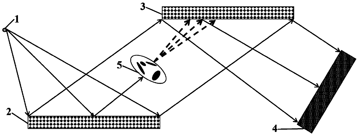
[0065] In this example, see figure 1 , setting an X-ray diffraction enhanced imaging system composed of X-ray source 1, monochromatic crystal 2, analysis crystal 3 and detector 4; as figure 1 As shown, the X-ray propagation direction is the Z axis; the X-ray source 1, the monochromatic crystal 2, the imaged object 5, the analysis crystal 3 and the detector 4 are arranged in sequence along the Z axis; then the X-ray based on the iterative algorithm The ray diffraction enhanced imaging method is carried out as follows:

[0066] Step 1. Set the relative position of each device, satisfying: 01 3 4 , where d 1 is the relative distance between the monochromatic crystal 2 and the X-ray source 1 along the Z axis, d 3 To analyze the relative distance between the crystal 3 and the X-ray source 1 along the Z axis; d 4 is the relative distance between the detector 4 and the X-ray source 1 along the Z-axis;

[0067] Step 2. Obtain background projection data:

[0068] Step 2.1, taking ...


PUM


Abstract

The invention discloses an X-ray diffraction enhanced imaging method based on an iterative algorithm. The method is applied to a diffraction enhanced imaging system composed of an X-ray source, a monochromatic crystal, an analysis crystal and a detector which are sequentially arranged in the X-ray propagation direction. X-rays are incident to the surface of the monochromatic crystal to be diffracted; emergent monochromatic collimated X-rays are diffracted after penetrating through an imaged object and entering the surface of the analysis crystal; after emergent X-rays are incident to the detector, the intensity of X-rays is detected and recorded by the detector, the normal of the diffraction surface of the analysis crystal is used as a rotating shaft, and the detector is used for recordingprojection data of the analysis crystal at three different angular positions, so that a proposed iterative algorithm is used for processing the projection data recorded by the detector, and absorption, refraction and scattering signals of an imaged object are obtained. The method can solve the problem of accurate extraction of the refraction signal and the scattering signal of the imaged object when the bias of the light intensity curve is not zero.

Description

technical field [0001] The invention relates to the field of X-ray imaging methods, in particular to an iterative algorithm-based X-ray diffraction enhanced imaging method. Background technique [0002] After more than 100 years of continuous development, X-ray imaging technology has been widely used in many fields such as clinical medical diagnosis and treatment, public safety inspection, and material science. For objects containing heavy metal elements, X-ray absorption contrast imaging technology can be used to obtain good imaging results. However, for objects mainly composed of low atomic number elements such as organic composite materials and human soft tissues, their attenuation to X-rays is very weak, and the image quality obtained by using absorption contrast imaging technology is very poor. In order to overcome this limitation, scientists have successively developed a series of multi-mode X-ray imaging methods as a powerful supplement to traditional absorption cont...

Claims


Application Information

Patent Timeline
IPC IPC(8): G01N23/207
CPCG01N23/207
Inventor 王志立陈恒
Owner HEFEI UNIV OF TECH
Why Eureka
Try Eureka