Supercharge Your Innovation With Domain-Expert AI Agents!

Tissue elasticity detection method, ultrasonic imaging equipment and computer storage medium

A technology of elastic detection and ultrasonic image, which is applied in the field of medical devices, can solve the problems of low detection efficiency, invalid detection results, complicated operation, etc., and achieve the effect of improving efficiency

Active Publication Date: 2020-09-08
SHENZHEN MINDRAY BIO MEDICAL ELECTRONICS CO LTD
View PDF9 Cites 0 Cited by
  • Summary
  • Abstract
  • Description
  • Claims
  • Application Information

AI Technical Summary

Problems solved by technology

[0003] In clinical practice, the existing technical scheme of instantaneous elastic detection requires the doctor to manually start each measurement, which is complicated to operate, and due to various factors such as patient movement, breathing, unstable operation techniques, and special state of the target tissue during the detection process, it may cause the detection The result is invalid, the doctor needs to manually supplement and start more measurements, and the detection efficiency is low

Method used


Image

  • Tissue elasticity detection method, ultrasonic imaging equipment and computer storage medium
  • Tissue elasticity detection method, ultrasonic imaging equipment and computer storage medium
  • Tissue elasticity detection method, ultrasonic imaging equipment and computer storage medium

Examples


Embodiment Construction

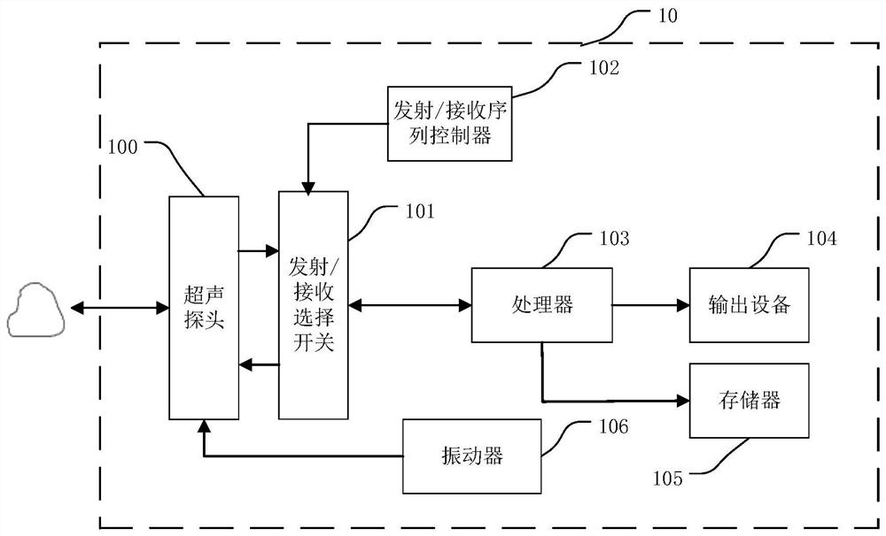
[0056] figure 1It is a schematic structural block diagram of the ultrasound imaging system 10 in the embodiment of the present application. The ultrasonic imaging system 10 may include an ultrasonic probe 100, a transmit / receive selection switch 101, a transmit / receive sequence controller 102, a processor 103, and an output device 104, wherein the output device may be a display, a speaker, or an indicator light etc., wherein the ultrasonic probe 100 can be a linear array probe, a convex array probe or a phased array probe, etc., and a suitable probe can be selected according to actual application scenarios. The transmit / receive sequence controller 102 can stimulate the ultrasound probe 100 to transmit ultrasound to the target tissue, and can also control the ultrasound probe 100 to receive ultrasound echoes returned from the target tissue, so as to obtain ultrasound echo signals / data. The processor 103 processes the ultrasound echo signal / data to obtain tissue-related paramet...


PUM


Abstract

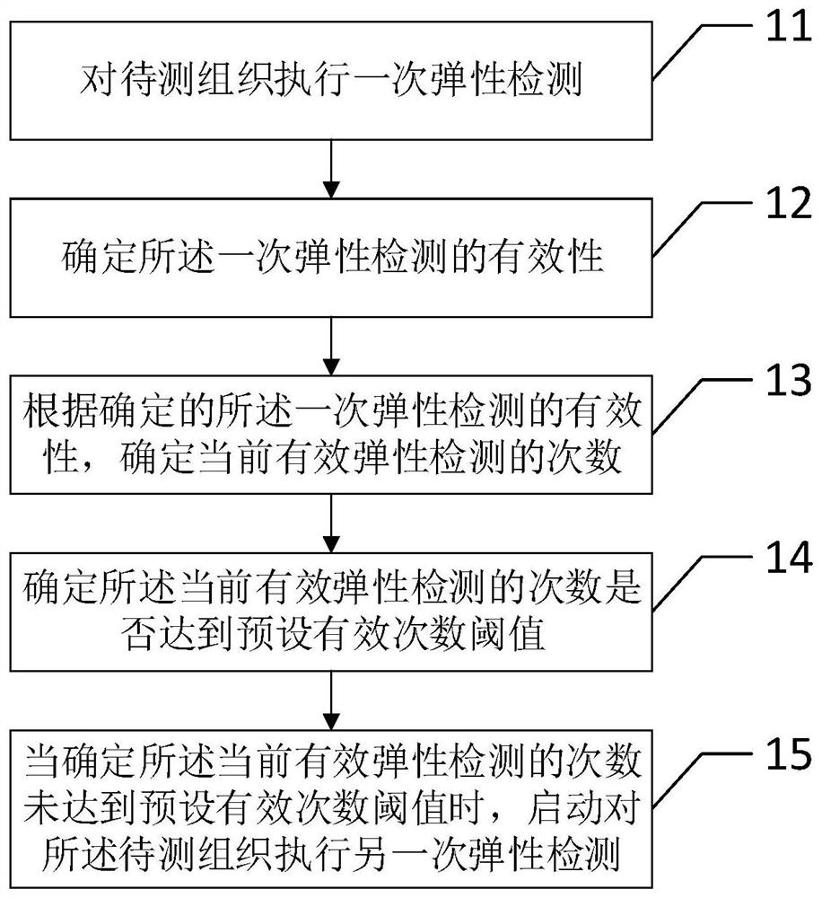
The invention provides a tissue elasticity detection method, ultrasonic imaging equipment and a computer storage medium. The tissue elasticity detection method comprises the following steps: performing primary elasticity detection on a to-be-detected tissue; determining effectiveness of the primary elasticity detection; determining the times of current effective elastic detection according to thedetermined effectiveness of the primary elastic detection; and determining whether the times of the current effective elastic detection reaches a preset effective time threshold, and starting execution of another time of elastic detection on the to-be-detected tissue when the times of the current effective elastic detection do not reach a preset effective time threshold. According to the method, repeated execution of elastic detection is automatically started to the to-be-detected tissue, and a user does not need to operate equipment for setting, so that usability and elastic detection efficiency are improved.

Description

technical field [0001] The present application relates to the field of medical devices, in particular to a method for detecting tissue elasticity, an ultrasonic imaging device and a computer storage medium. Background technique [0002] Transient Elastography (TE) plays an important role in the field of medical ultrasound imaging, one of which is the application in the field of liver fibrosis detection. Liver fibrosis is the pathological process of the development of various chronic liver diseases to liver cirrhosis. Elasticity technology reflects the degree of liver fibrosis by detecting the liver stiffness value. Compared with invasive pathological detection of liver biopsy, transient elasticity is non-invasive, simple, fast, easy to operate, repeatable, safe and well tolerated, and has been approved by WHO, AASLD, EASL, Chinese Medical Association for Liver Diseases, etc. It is recommended as an important means of clinical assessment of hepatitis B and C virus-related li...

Claims


Application Information

Patent Timeline
IPC IPC(8): A61B8/00
CPCA61B8/485A61B8/463
Inventor 李双双温博李金洋袁娟
Owner SHENZHEN MINDRAY BIO MEDICAL ELECTRONICS CO LTD
Features
  • R&D
  • Intellectual Property
  • Life Sciences
  • Materials
  • Tech Scout
  • Unparalleled Data Quality
  • Higher Quality Content
  • 60% Fewer Hallucinations
Learn More