Anti-cea antibody and application thereof, cell capable of secreting anti-cea antibody and preparation method thereof

An antibody and cell technology, applied in genetically modified cells, biochemical equipment and methods, cells modified by introducing foreign genetic material, etc., can solve the problem of poor specificity of anti-CEA antibodies

Active Publication Date: 2022-06-03
SHENZHEN YHLO BIOTECH +2
View PDF4 Cites 0 Cited by
  • Summary
  • Abstract
  • Description
  • Claims
  • Application Information

AI Technical Summary

Problems solved by technology

However, the existing anti-CEA antibodies have poor specificity and cannot meet the actual needs

Method used


Image

  • Anti-cea antibody and application thereof, cell capable of secreting anti-cea antibody and preparation method thereof
  • Anti-cea antibody and application thereof, cell capable of secreting anti-cea antibody and preparation method thereof
  • Anti-cea antibody and application thereof, cell capable of secreting anti-cea antibody and preparation method thereof

Examples


preparation example Construction

[0054] The method for preparing cells capable of secreting anti-CEA antibodies in one embodiment is characterized by comprising the following steps: using the mixture to transfect animal cells and culturing to obtain cells capable of secreting anti-CEA antibodies.

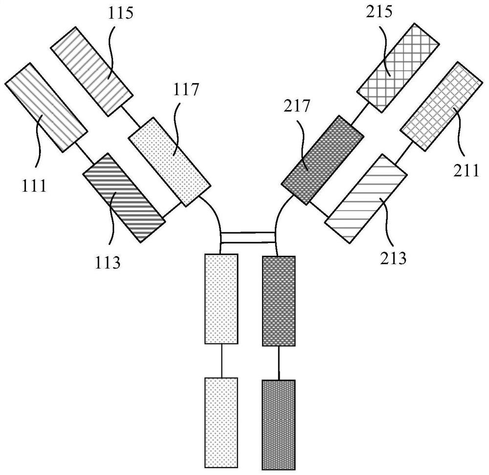
[0055] Wherein, the transfection composition comprises the first plasmid, the second plasmid and the transfection reagent. The first plasmid contains the coding sequence of the first light chain variable region and the coding sequence of the first heavy chain variable region, and the first light chain variable region The amino acid sequence of the region is shown in SEQ ID No.1, and the amino acid sequence of the first heavy chain variable region is shown in SEQ ID No.2. The second plasmid contains the coding sequence of the second light chain variable region and the coding sequence of the second heavy chain variable region, the amino acid sequence of the second light chain variable region is shown in SEQ ID No.3, a...

Embodiment 1

[0151] Monoclonal Antibody Preparation

[0152] Mix the immunogen and Freund's adjuvant in equal volumes and fully emulsify the mixture to obtain the mixture. Inject Balb / c mice with the mixture intraperitoneally to immunize Balb / c mice. A total of 3 immunizations, the interval between two adjacent immunizations For 15 days, the immunogen dose for each immunization was 25 μg / mouse.

[0153] After the immunization was completed, the spleen cells of the immunized mice were taken out, and 2.0×10 7 2.0×10 SP2 / 0 myeloma cells 8 Splenocytes of 1 immunized Balb / c mouse were mixed, centrifuged, the supernatant was discarded, slightly oscillated and mixed, placed in a water bath at 37°C, and 1 mL of 50% by volume PEG1500 aqueous solution was added dropwise within 90 seconds. Then add 20 mL of 1640 medium dropwise, centrifuge, discard the supernatant, wash once with PEG1500 aqueous solution and 1640 medium according to the above operation, centrifuge, discard the supernatant, and obta...

Embodiment 2

[0158] Antibody Titer Detection

[0159] The purified antibodies in Example 1 were diluted to concentrations of 3 μg / mL, 1 μg / mL, 0.33 μg / mL, 0.11 μg / mL, 0.036 μg / mL, and 0.012 μg / mL, and the antibody titer was detected by indirect ELISA method , ELISA test results are shown in Table 1. Wherein, the immunogen was diluted with CB buffer to a final concentration of 1 μg / mL to obtain a CB solution containing the immunogen. Add 100uL of CB solution containing immunogen to each well of the microtiter plate, and coat at 37°C for 3h. The microtiter plate was washed once with 0.01M PBST solution, and blocked by adding blocking solution (ie, PBS solution containing 20% ​​fetal bovine serum by mass percentage) at 37° C. for 2 hours. Wash the microtiter plate once with 0.01M PBST solution. Add 100uL of diluted antibody to each well (that is, dilute the above-mentioned anti-CEA monoclonal antibody 1000 times, 3000 times, 9000 times, 27000 times, 81000 times with ascites) and react at 3...


PUM


Abstract

The invention relates to an anti-CEA antibody and its application, a cell capable of secreting an anti-CEA antibody and a preparation method thereof. The anti-CEA antibody includes a first structural domain and a second structural domain. The first structural domain includes a first light chain variable region and a first heavy chain variable region. The amino acid sequence of the first light chain variable region is as SEQ ID No. 1, the amino acid sequence of the first heavy chain variable region is shown in SEQ ID No.2; the second structural domain includes the second light chain variable region and the second heavy chain variable region, and the second light chain variable region The amino acid sequence of the region is shown in SEQ ID No.3, and the amino acid sequence of the second heavy chain variable region is shown in SEQ ID No.4. The specificity of the above-mentioned anti-CEA antibody is better.

Description

technical field [0001] The invention relates to the field of biotechnology, in particular to an anti-CEA antibody and its application, a cell capable of secreting an anti-CEA antibody and a preparation method thereof. Background technique [0002] Cancer, also known as malignant tumor, can proliferate indefinitely and consume a large amount of nutrients in the patient's body. Cancer cells can also release a variety of toxins, causing a series of pathological symptoms in the human body. Cancer cells can also be transferred to the whole body. It grows and reproduces everywhere. The occurrence of cancer has brought great troubles to the lives of many patients, and even directly threatens the life and health of the body. For the treatment of cancer, the earlier it is found and treated, the better the treatment effect will be. [0003] Traditional tumor screening methods are mainly X-ray photography or detection of serum tumor markers, etc., but the irreversible damage caused b...

Claims


Application Information

Patent Timeline
Patent Type & Authority Patents(China)
IPC IPC(8): C07K16/46C12N15/85C12N5/10G01N33/574G01N33/68G01N33/558G01N33/543G01N33/535
CPCC07K16/3007C12N15/85C12N5/0686C12N5/0603G01N33/57473G01N33/57415G01N33/6854G01N33/558G01N33/543G01N33/535C07K2317/56C07K2317/52C07K2317/92C07K2317/14C07K2317/31C12N2510/02
Inventor 吴力强谢妮阎锡蕴刑智浩林晓涛钱纯亘胡鹍辉
Owner SHENZHEN YHLO BIOTECH
Why Eureka
Try Eureka