System and method for detecting abnormal tissue using vascular features

A vascular, abnormal technique, applied in the field of systems and methods for detecting abnormal tissue using vascular features, capable of addressing limitations, etc.

Pending Publication Date: 2020-10-20
KONINKLJIJKE PHILIPS NV
View PDF4 Cites 1 Cited by
  • Summary
  • Abstract
  • Description
  • Claims
  • Application Information

AI Technical Summary

Problems solved by technology

[0003] Moreover, minimally invasive surgery using surgical robotics and/or steerable d

Method used


Image

  • System and method for detecting abnormal tissue using vascular features
  • System and method for detecting abnormal tissue using vascular features
  • System and method for detecting abnormal tissue using vascular features

Examples


Embodiment Construction

[0018] Embodiments of the present invention will now be described more fully hereinafter with reference to the accompanying drawings in which embodiments of the invention are shown. However, this invention may be embodied in different forms and should not be construed as limited to the embodiments set forth herein. Rather, these embodiments are provided as teaching examples of the invention.

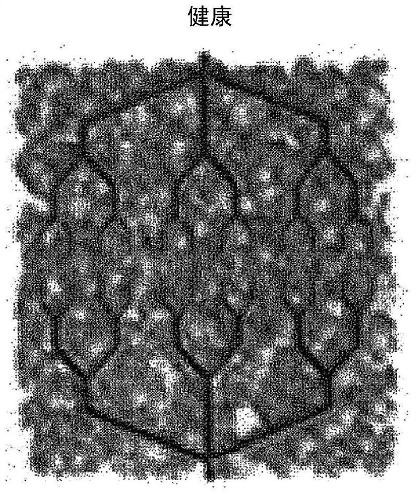
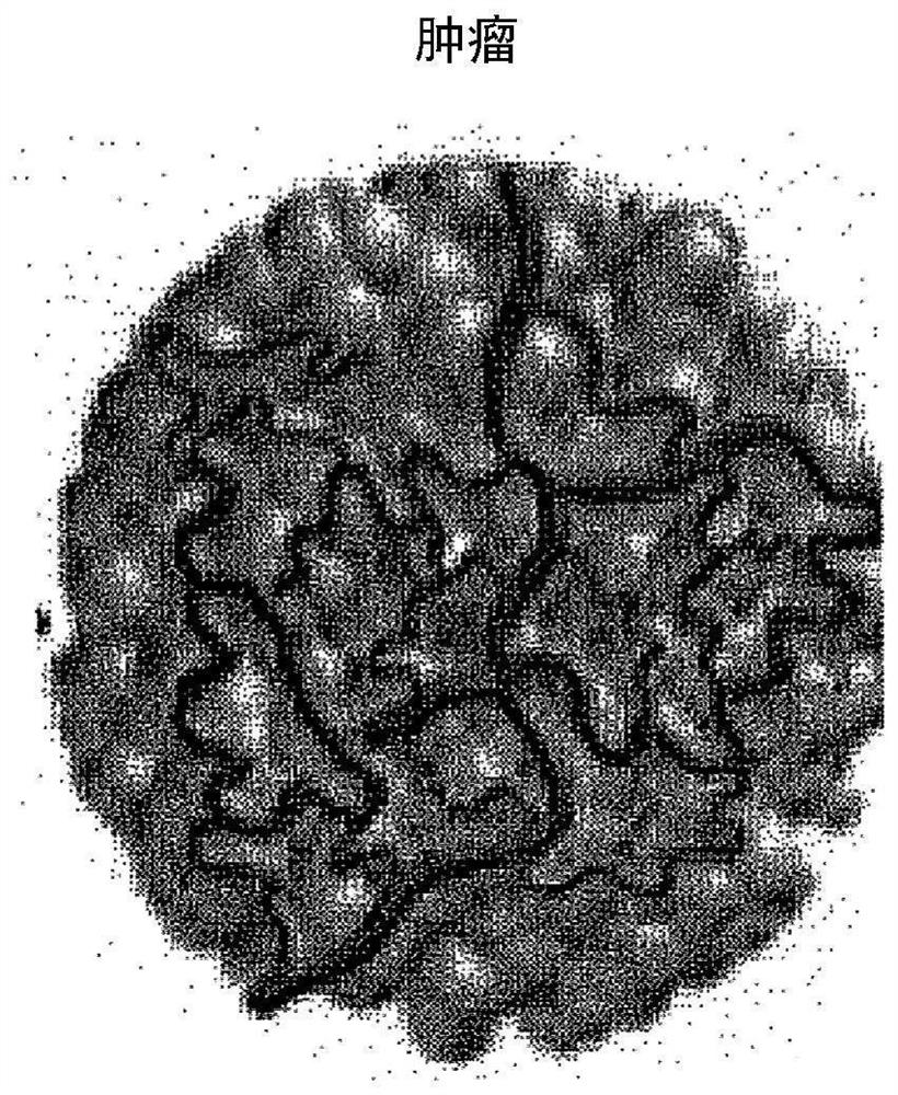
[0019] As noted above, intraoperative differentiation of abnormal tissue from normal tissue for resection and biopsy is a common challenge for surgeons performing various interventional procedures. According to various embodiments, vascular information extracted after creation of one or more Eulerian video magnifications indicative of vascular abnormalities or excessive vascular abnormalities may be used to assist a surgeon in differentiating abnormal tissue (e.g., a cancerous tumor) from normal tissue open. Additionally, various embodiments can be used to screen for suspected melanoma...


PUM


Abstract

An imaging system includes a camera, a display and a processor. The camera has color video acquisition capability, and is mounted to a distal end of an interventional instrument insertable within an object, the camera providing image frames for imaging vasculature of the object, each image frame including multiple pixels providing corresponding signals, respectively. The processor is programmed toreceive the signals; amplify variations in at least one of color and motion of the signals corresponding to the multiple pixels; determine spatial phase variability, frequency and signal characteristics of at least some of the amplified signals corresponding to the multiple pixels, respectively; identify pixels indicative of abnormal vascularity based on the spatial phase variability, frequency and/or signal characteristics; create a vascular map corresponding to each, where each vascularity map includes a portion of the object having the abnormal vascularity; and operate the display to display each vascularity map.

Description

Background technique [0001] The detection of tumors using vascularization is currently used in medical imaging. That is, tumors can be detected by analyzing blood vessels within the tumor because these tumor blood vessels are often dilated with irregular shapes, disordered connections, and leaky capillaries compared to normal blood vessels. For example, dynamic imaging of contrast agents injected into the venous system can be used to detect areas of increased vascularity, angiogenesis, and vascular leakage, all of which are indicative of cancerous tumors. However, cameras and optical imagers have not been used for cancerous tumor detection, even though video-linked surgical procedures are becoming more common. Minimally invasive surgical procedures often achieve improved patient outcomes. [0002] Distinguishing normal from abnormal tissue intraoperatively is often a challenge for the surgeon. For the unpalpable, often seen in brain cancers (eg, neurosurgery), lung cancers ...

Claims


Application Information

Patent Timeline
IPC IPC(8): G06T7/00
CPCG06T7/0012G06T2207/10072G06T2207/30101A61B1/00045A61B1/3137G02B27/017G06F3/011G06T2207/10016G06T2207/10024G06T2207/10068G06T2207/20056A61B1/000094
Inventor C·M·斯威舍
Owner KONINKLJIJKE PHILIPS NV
Why Eureka
Try Eureka